

FGF-dependent left-right asymmetry patterning in zebrafish is mediated by Ier2 and Fibp1
- PMID: 19164561
- PMCID: PMC2650137
- DOI: 10.1073/pnas.0812880106
FGF-dependent left-right asymmetry patterning in zebrafish is mediated by Ier2 and Fibp1
Abstract
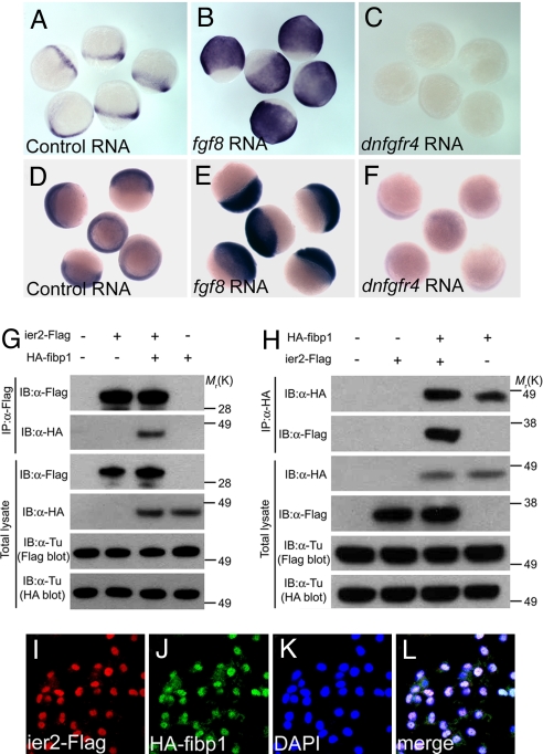
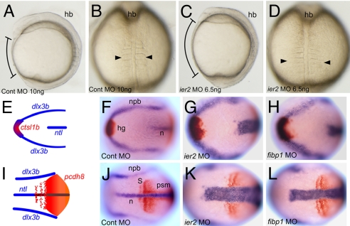
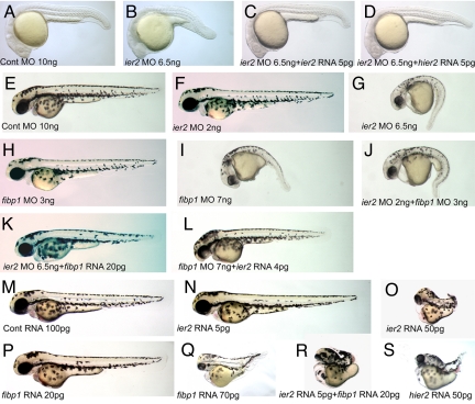
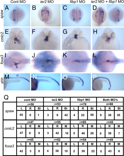
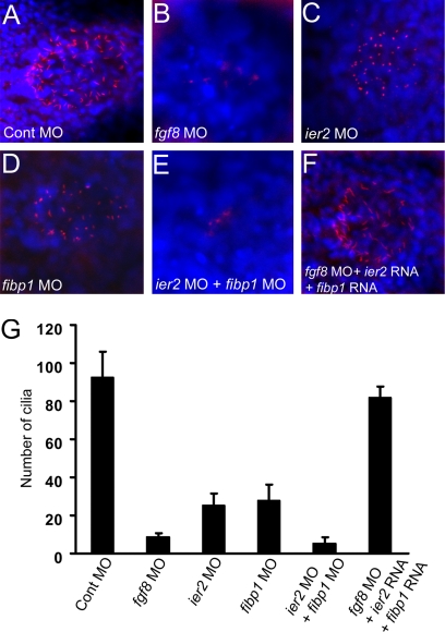
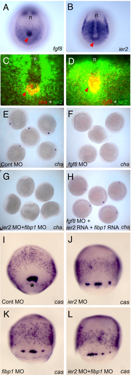
Establishment of left-right asymmetry in vertebrates requires nodal, Wnt-PCP and FGF signaling and involves ciliogenesis in a laterality organ. Effector genes through which FGF signaling affects laterality have not been described. We isolated the zebrafish ier2 and fibp1 genes as FGF target genes and show that their protein products interact. Knock down of these factors interferes with establishment of organ laterality and causes defective cilia formation in Kupffer's Vesicle, the zebrafish laterality organ. Cilia are also lost after suppression of FGF8, but can be rescued by injection of ier2 and fibp1 mRNA. We conclude that Ier2 and Fibp1 mediate FGF signaling in ciliogenesis in Kupffer's Vesicle and in the establishment of laterality in the zebrafish embryo.
Conflict of interest statement
The authors declare no conflict of interest.
Figures
Similar articles
-
ENC1-like integrates the retinoic acid/FGF signaling pathways to modulate ciliogenesis of Kupffer's Vesicle during zebrafish embryonic development.Dev Biol. 2013 Feb 1;374(1):85-95. doi: 10.1016/j.ydbio.2012.11.022. Epub 2012 Nov 30. Dev Biol. 2013. PMID: 23201577
-
Klf8 regulates left-right asymmetric patterning through modulation of Kupffer's vesicle morphogenesis and spaw expression.J Biomed Sci. 2017 Jul 17;24(1):45. doi: 10.1186/s12929-017-0351-y. J Biomed Sci. 2017. PMID: 28716076 Free PMC article.
-
Wnt/β-catenin signaling directly regulates Foxj1 expression and ciliogenesis in zebrafish Kupffer's vesicle.Development. 2012 Feb;139(3):514-24. doi: 10.1242/dev.071746. Epub 2011 Dec 21. Development. 2012. PMID: 22190638 Free PMC article.
-
Left-right asymmetry in zebrafish.Cell Mol Life Sci. 2012 Sep;69(18):3069-77. doi: 10.1007/s00018-012-0985-6. Epub 2012 Apr 19. Cell Mol Life Sci. 2012. PMID: 22527718 Free PMC article. Review.
-
Fish and frogs: models for vertebrate cilia signaling.Front Biosci. 2008 Jan 1;13:1866-80. doi: 10.2741/2806. Front Biosci. 2008. PMID: 17981674 Free PMC article. Review.
Cited by
-
FGF signalling: diverse roles during early vertebrate embryogenesis.Development. 2010 Nov;137(22):3731-42. doi: 10.1242/dev.037689. Development. 2010. PMID: 20978071 Free PMC article. Review.
-
Fuz mutant mice reveal shared mechanisms between ciliopathies and FGF-related syndromes.Dev Cell. 2013 Jun 24;25(6):623-35. doi: 10.1016/j.devcel.2013.05.021. Dev Cell. 2013. PMID: 23806618 Free PMC article.
-
Swimming toward solutions: Using fish and frogs as models for understanding RASopathies.Birth Defects Res. 2020 Jun;112(10):749-765. doi: 10.1002/bdr2.1707. Epub 2020 Jun 7. Birth Defects Res. 2020. PMID: 32506834 Free PMC article. Review.
-
The ciliary baton: orchestrating neural crest cell development.Curr Top Dev Biol. 2015;111:97-134. doi: 10.1016/bs.ctdb.2014.11.004. Epub 2015 Jan 22. Curr Top Dev Biol. 2015. PMID: 25662259 Free PMC article. Review.
-
Origin, form and function of extraembryonic structures in teleost fishes.Philos Trans R Soc Lond B Biol Sci. 2022 Dec 5;377(1865):20210264. doi: 10.1098/rstb.2021.0264. Epub 2022 Oct 17. Philos Trans R Soc Lond B Biol Sci. 2022. PMID: 36252221 Free PMC article. Review.
References
-
- Bisgrove BW, Morelli SH, Yost HJ. Genetics of human laterality disorders: Insights from vertebrate model systems. Annu Rev Genomics Hum Genet. 2003;4:1–32. - PubMed
-
- Hamada H, Meno C, Watanabe D, Saijoh Y. Establishment of vertebrate left–right asymmetry. Nat Rev Genet. 2002;3:103–113. - PubMed
-
- Raya A, Belmonte JC. Left-right asymmetry in the vertebrate embryo: From early information to higher-level integration. Nat Rev Genet. 2006;7:283–293. - PubMed
-
- Capdevila J, et al. Mechanisms of left–right determination in vertebrates. Cell. 2000;101:9–21. - PubMed
-
- Speder P, Petzoldt A, Suzanne M, Noselli S. Strategies to establish left/right asymmetry in vertebrates and invertebrates. Curr Opin Genet Dev. 2007;17:351–358. - PubMed
Publication types
MeSH terms
Substances
Grants and funding
LinkOut - more resources
Full Text Sources
Molecular Biology Databases
