. 2009 Jan 27;106(4):1239-44.
doi: 10.1073/pnas.0810794106. Epub 2009 Jan 21.

RodZ, a component of the bacterial core morphogenic apparatus

Affiliations

RodZ, a component of the bacterial core morphogenic apparatus

S Anisah Alyahya et al. Proc Natl Acad Sci U S A. .

Abstract

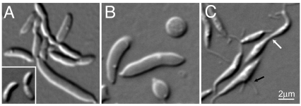
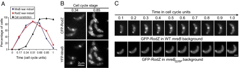
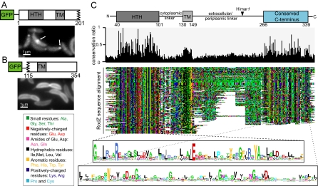
The molecular basis of bacterial cell morphogenesis remains largely an open question. Here we discover a morphogenic protein, RodZ, which is widely conserved across the bacterial kingdom. In Caulobacter crescentus, RodZ is essential for viability and is involved in all aspects of this organism's complex morphology. Depletion or over-production of RodZ results in grossly misshapen cells with stalk defects. RodZ exhibits a localization pattern during the cell cycle corresponding to sites of active peptidoglycan synthesis. The temporal transition of RodZ between patchy/helical and mid-cell localization mimics and depends on the actin-like MreB cytoskeleton. In Escherichia coli, an organism with a distinct mode of growth and MreB localization dynamics, RodZ follows MreB and retains its crucial role in cell morphogenesis, demonstrating conservation of function. Genomic analysis shows that RodZ represents an ancient function unique to bacteria. Multiple sequence alignment of 143 RodZ sequences from species across bacterial phyla identifies an N-terminal cytoplasmic domain with a helix-turn-helix motif, a transmembrane sequence, and a previously unidentified, conserved periplasmic or extracellular C-terminal domain. Both the N- and C-terminal domains are important for function, with the N-terminal domain containing localization determinants. This study uncovers a key missing player in the cytoskeleton-based growth machinery enabling heritable and defined cellular forms in bacteria.


Conflict of interest statement

The authors declare no conflict of interest.

Figures

Fig. 1.
Fig. 1.
Identification of a cell morphogenic gene. (A) DIC micrograph of CB15N rodZ::Himar1 mutant cells. Inset shows wild-type cells for comparison. (B) Depletion of RodZ in CJW2747 cells (CB15N rodZ::Ω xylX::pXrodZ) was initiated by substituting xylose (inducer) in PYE with glucose to halt rodZ expression. The DIC image was taken after 28 h of depletion. (C) Overproduction of RodZ in CJW2158 cells (CB15N/pJS14PxylrodZ) was initiated by addition of xylose. The DIC image was taken after 20 h of over-production. White and black arrows show thin cellular connections between cells and a medial bulge with 2 stalks, respectively.
Fig. 2.
Fig. 2.
RodZ localizes to areas of peptidoglycan growth and depends on FtsZ for its medial localization. (A) CJW2867 cells (CB15N xylX::pXCFPN-1rodZ vanA::pMT400) producing CFP-RodZ under Pxyl and FtsZ-YFP under Pvan were grown in the presence of vanillic acid and xylose for 2 h before synchronization. Synchronized swarmer cells were resuspended in liquid medium containing vanillate and xylose, and samples were taken at 15-min intervals for microscopy. Under these conditions, the cell cycle takes about 90 min. Time is annotated in cell cycle units; 0 immediately follows synchronization and 1 represents cell separation. Arrows show stalks. (Scale bar, 2 μm.) (B) CJW2907 cells (CB15N rodZ::pHL23Pxylgfp-rodZ ftsZ::pVMCS-6FtsZ5′) grown in vanillate-containing medium were washed and resuspended in vanillate-free medium containing xylose to induce GFP-RodZ synthesis while depleting FtsZ. Cells were imaged 3 h later.
Fig. 3.
Fig. 3.
RodZ localization depends on MreB. (A) Quantitative analysis of cells with medial localization of YFP-MreB (blue), with medial localization of CFP-RodZ (red) and with discernible cell constrictions (black) during the course of the cell cycle. Results obtained from 2 separate time-course experiments in which over 100 cells were considered at each time point. Strain CJW2866 (CB15N rodZ::cfp-rodZ xylX::pXYFP-mreB) was grown with xylose for 2 h before synchronization to induce the expression of yfp-mreB. Synchronized swarmer cells were resuspended in PYE with xylose and samples were taken at regular time intervals converted to cell-cycle units to examine the localization of YFP-MreB and CFP-RodZ over the cell cycle. (B) Representative images of YFP-MreB and CFP-RodZ localizations from the time-course experiments described in (A). See Fig. S3 for the whole time-course sequence. (C) Time-lapse microscopy of GFP-RodZ in an mreB wild-type background (CJW2748; CB15N xylX::pXGFPN-2rodZ) or in an mreBQ26P mutant background (CJW2767; CB15N mreBQ26P xylX::pXGFPN-2rodZ) over the cell cycle. GFP-RodZ synthesis was induced with xylose for 2 h before microscopy. The agarose-padded slide contained xylose. (Scale bar, 1 μm.)
Fig. 4.
Fig. 4.
Mutagenesis and multiple sequence alignment identify regions and residues of RodZ important for its structure, function, and localization. (A) Fluorescence micrograph of CJW2936 cells (CB15N xylX::pXGFPN-2rodZ1–201) preinduced with xylose for 2 h to synthesize GFP-RodZ1–201 (in a wild-type rodZ background). White arrows indicate polar and medial localizations. (B) Fluorescence micrograph of CJW2869 cells (CB15N xylX::pXGFPN-2rodZ115–354) preinduced with xylose for 2 h to synthesize GFP-RodZ115–354 (in a wild-type rodZ background). (C) Colored dotplot showing alignment of 143 RodZ sequences and identifying conserved regions. (Top) Conservation histogram ranging from 0 (no conservation) to 1 (100% conservation). (Bottom) logos showing consensus sequences for the HTH and C-terminal domains; letter height is proportional to the degree of conservation.
Fig. 5.
Fig. 5.
The cell shape-determining function and the MreB-dependent localization of RodZ is conserved in E. coli. (A) DIC images of rodZ knockout cells (CJW2910; MC1000 ΔrodZEc::Kan) from log-phase (Left) and stationary phase (Right) populations. The insets show wild-type E. coli MC1000 cells for comparison. (B) Fluorescent and DIC images of GFP-RodZEc-overproducing cells (CJW2912; MC1000/pBAD18gfprodZEc) after 10 h of growth in arabinose. (C) Fluorescence image of cells producing GFP-RodZEc (CJW2871; MC1000/pBAD33gfprodZEc). After a 10-min arabinose induction, glucose was added for another 20 min before microscopy. (D) CJW2911 cells (MC1000/pBAD33gfprodZEc/pLE7) were treated with isopropyl-beta-D-thiogalactopyranoside (IPTG) for 2 h (to induce YFP-MreB synthesis) and cephalexin for 1.5 h before microscopy. Induction of GFP-RodZEc was achieved as in (C). (E) GFP-RodZEc localization in ΔmreB cells (CJW2913; PB103mreB<>frt/pFB112/pBAD33gfprodZEc). Induction of GFP-RodZEc was achieved as in (C). (F) YFP-MreB localization in a rodZ knockout background (CJW2946; MC1000ΔrodZEc::Kan/pLE7) after 2 h of growth in the presence of IPTG to induce YFP-MreB synthesis.
Fig. 6.
Fig. 6.
Phlyogenetic tree of bacterial species showing the presence of RodZ (red), MreB (blue), MreC (cyan), MreD (purple), adjacency between rodZ and gcpE (green), and adjacency between rodZ and pgsA (black). Subtrees indicate phyla, except for the proteobacterial phylum, which is further subdivided by class. See Fig. S5 for a more detailed tree.

References

    1. Shih YL, Rothfield L. The bacterial cytoskeleton. Microbiol Mol Biol Rev. 2006;70:729–754. - PMC - PubMed
    1. Pichoff S, Lutkenhaus J. Overview of cell shape: cytoskeletons shape bacterial cells. Curr Opin Microbiol. 2007;10:601–605. - PMC - PubMed
    1. Osborn MJ, Rothfield L. Cell shape determination in Escherichia coli. Curr Opin Microbiol. 2007;10:606–610. - PubMed
    1. Jones LJ, Carballido-Lopez R, Errington J. Control of cell shape in bacteria: helical, actin-like filaments in Bacillus subtilis. Cell. 2001;104:913–922. - PubMed
    1. Wachi M, et al. Mutant isolation and molecular cloning of mre genes, which determine cell shape, sensitivity to mecillinam, and amount of penicillin-binding proteins in Escherichia coli. J Bacteriol. 1987;169:4935–4940. - PMC - PubMed

Publication types