. 2009 Feb 3;106(5):1638-41.
doi: 10.1073/pnas.0811682106. Epub 2009 Jan 21.

Targeted disruption of the Cl-/HCO3- exchanger Ae2 results in osteopetrosis in mice

Affiliations

Targeted disruption of the Cl-/HCO3- exchanger Ae2 results in osteopetrosis in mice

Kaj Josephsen et al. Proc Natl Acad Sci U S A. .

Abstract

Osteoclasts are multinucleated bone-resorbing cells responsible for constant remodeling of bone tissue and for maintaining calcium homeostasis. The osteoclast creates an enclosed space, a lacuna, between their ruffled border membrane and the mineralized bone. They extrude H(+) and Cl(-) into these lacunae by the combined action of vesicular H(+)-ATPases and ClC-7 exchangers to dissolve the hydroxyapatite of bone matrix. Along with intracellular production of H(+) and HCO(3)(-) by carbonic anhydrase II, the H(+)-ATPases and ClC-7 exchangers seems prerequisite for bone resorption, because genetic disruption of either of these proteins leads to osteopetrosis. We aimed to complete the molecular model for lacunar acidification, hypothesizing that a HCO(3)(-) extruding and Cl(-) loading anion exchange protein (Ae) would be necessary to sustain bone resorption. The Ae proteins can provide both intracellular pH neutrality and serve as cellular entry mechanism for Cl(-) during bone resorption. Immunohistochemistry revealed that Ae2 is exclusively expressed at the contra-lacunar plasma membrane domain of mouse osteoclast. Severe osteopetrosis was encountered in Ae2 knockout (Ae2-/-) mice where the skeletal development was impaired with a higher diffuse radio-density on x-ray examination and the bone marrow cavity was occupied by irregular bone speculae. Furthermore, osteoclasts in Ae2-/- mice were dramatically enlarged and fail to form the normal ruffled border facing the lacunae. Thus, Ae2 is likely to be an essential component of the bone resorption mechanism in osteoclasts.


Conflict of interest statement

The authors declare no conflict of interest.

Figures

Fig. 1.
Fig. 1.
Immunolocalization of Ae2 in mouse and rat osteoclasts in paraffin sections of decalcified tissue blocks. (A) Laser scanning confocal microscopy demonstrated intense immunostaining of the curved contra-lacunar surfaces in osteoclasts from the skull base of Ae2+/+ mice (green fluorescence, arrows). No immunostaining was seen corresponding to the ruffled border (rb). (B) Osteoclasts from the same area in knockout mice (Ae2−/−) showed no surface labeling (arrows). Nuclei are shown in red, many of which belong to bone marrow cells (bm). Black background represents bone tissue (asterisks). (C and D) Rat osteoclasts (oc) attached to alveolar bone (ab) surrounding the maxillary incisor also showed labeling of the contra-lacunar surface domain and unlabeled ruffled borders (rb). cyt, osteocytes; ct, connective tissue of periodontal membrane; gld, glandular tissue of the lateral nasal gland cupping the outer aspect of the alveolar bone also shows immunolocalization of Ae2. Rat, Ae2+/+, and Ae2−/− mouse osteoclasts also displayed a weak cytoplasmic labeling. (Scale bars, 20 μm.)
Fig. 2.
Fig. 2.
Ae2-deficient mice develop osteopetrosis. (A and B) X-ray images of half heads from 15-day-old mice demonstrated an increase in bone density in knockout mice (Ae2−/−) compared with controls (Ae2+/+) especially at the skull base (asterisk). ui, upper incisor; li, lower incisor; mr, molar tooth region. (Scale bars, 5 mm.) (C and D) Sagittal sections from methyl methacrylate embedded, undecalcified tissue blocks of the base of the skull from Ae2+/+ and Ae2−/− mice stained with Goldner's trichrome. The wide bone marrow cavity (bm) in Ae2+/+ mice filled with hematopoietic cells was in Ae2−/− mice almost obliterated by interwoven endochondral bone trabeculae. sos, cartilage plate of spheno-occipital synchondrosis; arrows, osteoclasts. (Scale bars, 200 μm.)
Fig. 3.
Fig. 3.
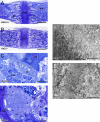
Epon sections of decalcified bone tissue from the skull base of Ae2+/+ and Ae2−/− mice stained with toluidine blue. Osteoclasts in Ae2−/− mice are morphologically distinct from those of Ae2+/+ mice. (A) In Ae2+/+ mice, the endochondral bone formation at the skull base was characterized by a smaller number of bone trabeculae (bt) with narrow remnants of cartilage matrix. Large medullary cavities (bm) with osteoclasts (arrows) were seen on each side of the cartilage plate of the spheno-occipital synchondrosis (sos). (B) In Ae2−/− mice, the bone trabeculae were interweaving and contained wide plates of cartilage matrix. The medullary cavity was reduced to narrow compartments occupied by voluminous osteoclasts (arrows). sos, spheno-occipital synchondrosis. (Scale bars, 200 μm.) (C) Osteoclasts (oc) from Ae2+/+ mice showed a distinct ruffled border (arrows) apposed to the bone. bt, bone trabecula with cartilage matrix (ct); bmc, bone marrow cells. (D) In Ae2−/− mice, osteoclasts (oc, inside dotted boundary line) were expanded in size and did not form distinct ruffled borders (arrows). Trabeculae of cartilage matrix plates (ct) were bordered by a thin layer of bone matrix (bo) having a basophilic surface zone. (Scale bars, 10 μm.) (E) Electron micrograph of the resorptive surface area in an osteoclast (oc) from Ae2+/+ mice reveals the complex folding of the resorbing surface into a ruffed border (asterisks) with deep invaginations containing collagen fibrils from the bone matrix (bo). (F) In Ae2−/− mice, the cell surface of osteoclasts (oc) apposed to bone (arrows) was smooth without membrane invaginations (arrows) but demonstrated coated endocytosis vesicles (cv). A finely textured granular layer (gl) was present between the osteoclast membrane and a loosely structured collagen matrix of the bone (bo). [Scale bars, 1 μm (E) and 500 nm (F).]
Fig. 4.
Fig. 4.
Diagram of an osteoclast indicating the processes involved in acidification of the resorptive lacuna. The osteoclast is attached on bone with a tight circumferential sealing zone (Sz) that divides the cell surface into two separate domains, the resorptive surface (Rs), folded into a ruffled border within the sealing zone, and the contra-lacunar surface (Cs). Intracellular H+ and HCO3 are formed from H2O and CO2 catalyzed by carbonic anhydrase II (CA II). H+ is transported across the ruffled border into the resorption lacuna by a vacuolar-type H+-ATPase. Cl ions follow H+ to the lacunae through the H+/Cl exchangers (ClC-7) and maintain electroneutrality. Excess HCO3 would, in time, cause cytoplasmic alkalinization and is removed via a contra-lacunar Cl/HCO3 exchange by Ae2, which also provides the cells with Cl for secretion into the lacunar space.

References

    1. Tolar J, Teitelbaum SL, Orchard PJ. Osteopetrosis. N Engl J Med. 2004;351:2839–2849. - PubMed
    1. Blair HC, Teitelbaum SL, Ghiselli R, Gluck S. Osteoclastic bone resorption by a polarized vacuolar proton pump. Science. 1989;245:855–857. - PubMed
    1. Vaananen HK, et al. Evidence for the presence of a proton pump of the vacuolar H(+)-ATPase type in the ruffled borders of osteoclasts. J Cell Biol. 1990;111:1305–1311. - PMC - PubMed
    1. Kornak U, et al. Loss of the ClC-7 chloride channel leads to osteopetrosis in mice and man. Cell. 2001;104:205–215. - PubMed
    1. Silver IA, Murrills RJ, Etherington DJ. Microelectrode studies on the acid microenvironment beneath adherent macrophages and osteoclasts. Exp Cell Res. 1988;175:266–276. - PubMed

Publication types

LinkOut - more resources