. 2009 Jan 27;106(4):1175-80.
doi: 10.1073/pnas.0806936106. Epub 2009 Jan 21.

Image-guided, noninvasive, spatiotemporal control of gene expression

Affiliations

Image-guided, noninvasive, spatiotemporal control of gene expression

Roel Deckers et al. Proc Natl Acad Sci U S A. .

Abstract

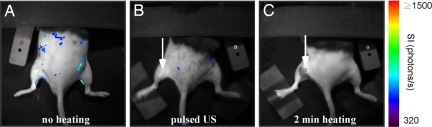
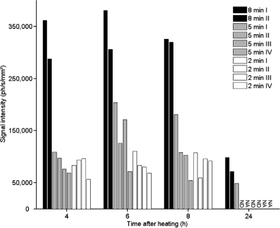
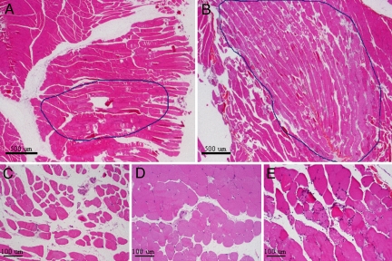
Spatiotemporal control of transgene expression is of paramount importance in gene therapy. Here, we demonstrate the use of magnetic resonance temperature imaging (MRI)-guided, high-intensity focused ultrasound (HIFU) in combination with a heat-inducible promoter [heat shock protein 70 (HSP70)] for the in vivo spatiotemporal control of transgene activation. Local gene activation induced by moderate hyperthermia in a transgenic mouse expressing luciferase under the control of the HSP70 promoter showed a high similarity between the local temperature distribution in vivo and the region emitting light. Modulation of gene expression is possible by changing temperature, duration, and location of regional heating. Mild heating protocols (2 min at 43 degrees C) causing no tissue damage were sufficient for significant gene activation. The HSP70 promoter was shown to be induced by the local temperature increase and not by the mechanical effects of ultrasound. Therefore, the combination of MRI-guided HIFU heating and transgenes under control of heat-inducible HSP promoter provides a direct, noninvasive, spatial control of gene expression via local hyperthermia.


Conflict of interest statement

The authors declare no conflict of interest.

Figures

Fig. 1.
Fig. 1.
HSP70 promoter activity in bone marrow cells 5 h after heating at different temperatures (42 °C, 43 °C, 44 °C, and 45 °C) and for different durations (0, 1, 2, 4, 8, 16, and 32 min). Promoter activity was measured as emitted light by luciferase by using an in vitro enzymatic assay.
Fig. 2.
Fig. 2.
Typical time course of the temperature evolution during a heating experiment (A), with the target temperature (in black) and the measured temperature at the focal point (in red). (B) MRI temperature map (color-coded) superimposed on an anatomical MRI image (grayscale) of the mouse leg.
Fig. 3.
Fig. 3.
MRI temperature maps of 2 mice heated with the same protocol, i.e., 8 min of heating at 43 °C at the focal point (A and B). The colors in A and B show the local temperature distribution in the mouse leg in the range of 38 °C (blue) to 42 °C and higher (red). (C and D) Bioluminescence images of the same 2 mice taken 6 h after the heating protocol. The colors in C and D show the light intensity measured with an optical CCD camera in the range of 320 photons per second (blue) to 2,000 photons per second and more (red).
Fig. 4.
Fig. 4.
Bioluminescence images taken before (A) and 6 h after (B) depositing 125 J of energy with pulsed ultrasound, and 6 h after heating at 43 °C for 2 min (C). The arrows indicate the locations of HIFU application. The color coding represents the light intensity measured with the CCD camera in photons per second.
Fig. 5.
Fig. 5.
Time course of gene expression in vivo upon hyperthermia. The leg muscle of transgenic mice was heated at 43 °C for 2 min (n = 4), 5 min (n = 4), or 8 min (n = 2). The mean light intensity is reported in photons per second per mm2. Light emitted by luciferase was measured at 4, 6, 8, and 24 h after heating. Light emission below the quantification threshold was marked as ND (not detectable), and measurements not performed are marked as NA (not available).
Fig. 6.
Fig. 6.
H&E-stained histology sections of excised muscle tissue 24 h after heating at 43 °C for 5 min (A and C) and 8 min (B and E). (D) After 5 min of heating, a modest variability in fiber diameters and interstitial edema was observed compared with the unheated muscle. The muscular damage was more extensive after 8 min of heating, including necrotic fibers and inflammatory infiltrates. (Magnification: A and B, 2×; C–E, 10×.)

Similar articles

Cited by

References

    1. Vilaboa N, Voellmy R. Regulatable gene expression systems for gene therapy. Curr Gene Ther. 2006;6:421–438. - PubMed
    1. Gorski K, Carneiro M, Schibler U. Tissue-specific in vitro transcription from the mouse albumin promoter. Cell. 1986;47:767–776. - PubMed
    1. Melo LG, et al. Endothelium-targeted gene and cell-based therapies for cardiovascular disease. Arterioscler Thromb Vasc Biol. 2004;24:1761–1774. - PubMed
    1. Rendahl KG, et al. Regulation of gene expression in vivo following transduction by two separate rAAV vectors. Nat Biotechnol. 1998;16:757–761. - PubMed
    1. Wang Y, DeMayo FJ, Tsai SY, O'Malley BW. Ligand-inducible and liver-specific target gene expression in transgenic mice. Nat Biotechnol. 1997;15:239–243. - PubMed

Publication types

Substances