. 2009 Feb 17;106(7):2236-41.
doi: 10.1073/pnas.0803202106. Epub 2009 Jan 21.

An essential role for Radar (Gdf6a) in inducing dorsal fate in the zebrafish retina

Affiliations

An essential role for Radar (Gdf6a) in inducing dorsal fate in the zebrafish retina

Nathan J Gosse et al. Proc Natl Acad Sci U S A. .

Abstract

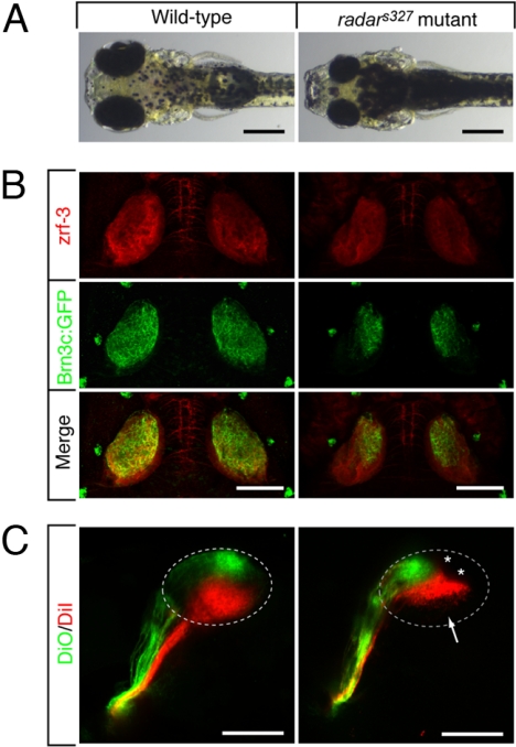
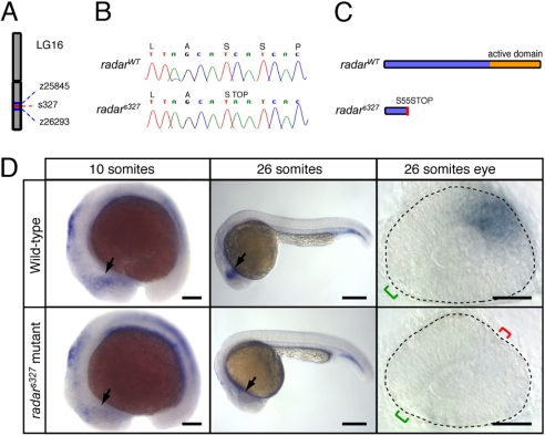
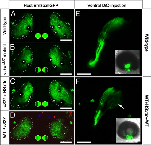
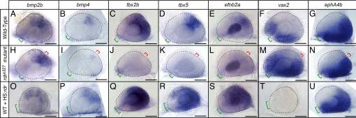
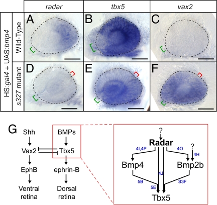
Retinal ganglion cells form orderly topographic connections with the tectum, establishing a continuous neural representation of visual space. Mapping along the dorsal-ventral axis requires interactions between EphB and ephrin-B cell-surface molecules expressed as countergradients in both retina and tectum. We have discovered that the diffusible TGFss-related factor Radar (Gdf6a) is necessary and sufficient for activation of dorsal markers, such as Bmp4, Tbx5, Tbx2b, and Ephrin-B2, and suppression of the ventral marker Vax2 in the zebrafish retina. Radar mutant axons innervate only the dorsal half of the tectum, where they form a compressed retinotectal map. Wild-type cells transplanted into the dorsal retina are able to rescue the dorsal identity of nearby mutant cells. Moreover, Radar overexpression "dorsalizes" retinal ganglion cell identity in the ventral retina. We conclude that Radar is near the top of a signaling cascade that establishes dorsal-ventral positional information in the retina and controls the formation of the retinotectal map.


Conflict of interest statement

The authors declare no conflict of interest.

Figures

Fig. 1.
Fig. 1.
Morphological and retinotectal phenotypes of radars327. (A) Zebrafish radars327 mutants have small eyes and appear dark, because of a VBA defect (see text). Dorsal brightfield views of 7 dpf WT sibling and homozygous radars327 mutant larvae. (B) radars327 mutants lack ventral innervation of the optic tectum. Dorsal confocal projections of 7 dpf larvae show that innervating RGC axons (expressing Brn3c:mGFP) are confined to the dorsal tectum in the mutant. Costaining with a neuropil marker (zrf-3 antibody) reveals that the size of the tectum is similar in WT and mutant. (C) radars327 mutants have a compressed dorsal–ventral retinotectal map. Fixed WT and radars327 eyes (7 dpf) were injected with DiO (ventrally) and DiI (dorsally). Lateral confocal projections are shown. Arrow highlights ventral tectal region not innervated by RGCs in radars327; asterisks show positions of skin melanophores. (Scale bars: 300 μm in A, 100 μm in B and C.)
Fig. 2.
Fig. 2.
Positional cloning and expression pattern of radar. (A) s327 maps to chromosome 16 between z25845 (2.3 cM) and z26293 (0.7 cM). (B) Sequencing of WT and s327 cDNA reveals a single C-to-A substitution in position 164 of the radar ORF, resulting in a premature stop codon. (C) Predicted translated peptides arising from radarWT and radars327. The mutation is predicted to result in a truncated protein, lacking the mature signaling domain. (D) Whole-mount in situ hybridization shows a restricted pattern of radar expression in WT embryos. radar mRNA is largely absent from the retina of radars327 mutants at all stages. In WT, expression is evident in the distal optic vesicle of WT embryos at 10 somites (arrow). At 26 somites, radar is expressed dorsally, opposite of the optic fissure (green bracket). Note ectopic fissures (red bracket) in radars327 mutants. (Scale bars: 150 μm for 10 somites, 250 μm for 26 somites, 50 μm for dissected 26 somite eyes.)
Fig. 3.
Fig. 3.
Cell–cell signaling through Radar is sufficient for ventral tectum innervation. (A–D) Rescue experiment. Brn3c:mGFP-labeled retinotectal projections were imaged in vivo at 7 dpf. The tectal neuropil is outlined with a dashed line. Green-filled circles summarize dorsal and ventral tectum innervation results. (A) WT tecta show full innervation. (B) radars327 mutants lack ventral innervation. (C) radarWT expression from heatshock-promoter rescues the retinotectal phenotype of the mutant. In the case shown, only one side was rescued. PCR-based genotyping confirmed that only the rescued eye contained hsp70:radarWT; the other eye had likely not received the injected plasmid because of the mosaicism inherent in transient transgenesis. (D) The radar gene acts cell-nonautonomously in the retina. WT cells transplanted into radars327 host embryos are sufficient to rescue ventral innervation. Only the host carried the Brn3c:mGFP transgene. Donor-derived cells (blue arrowheads) were labeled with rhodamine dextran, and do not contribute to the tectum. (E and F) Gain-of-function experiment. DiO was injected into the ventral retina, and its labeling pattern was imaged from a lateral view. Insets show injected eye. In normal WT larvae (E), ventral RGCs project exclusively to the dorsal tectum. In chimeric WT larvae (F) that have received a transplant of WT cells carrying the hsp70:radarWT construct, some ventral axons ectopically innervate the ventral tectum (arrow). Asterisks (in A–D) show positions of skin melanophores. (Scale bars, 100 μm.)
Fig. 4.
Fig. 4.
Radar signaling affects known determinants of dorsal–ventral retinal patterning. Expression patterns are visualized by whole-mount in situ hybridization of eyes from 26-somite embryos. Retinal markers in WT (A–G), radars327 mutants (H--N), and WT overexpressing hsp70:radarWT (O--U). In radars327 mutants, retinal bmp2b expression remained normal, bmp4 expression was severely reduced, tbx2b, tbx5, and efnb2a expression was absent, and vax2 expression was expanded dorsally. In radar-overexpressing embryos, bmp2b, bmp4, tbx2b, tbx5 and efnb2a expression were expanded ventrally, and vax2 was lost. ephA4b was unaffected by either loss or gain of radar function. Eyes overexpressing radar were often small and failed to close at the optic fissure. Neural retina is outlined with dashed line; green bracket identifies optic fissure location and size; red bracket indicates location of ectopic fissures in mutants. (Scale bars, 50 μm.)
Fig. 5.
Fig. 5.
Overexpression of bmp4 requires radar to alter dorsal–ventral patterning. (A--F) Expression patterns visualized by whole-mount in situ hybridization of eyes from 26-somite embryos. Neural retina is outlined with dashed line; green bracket identifies optic fissure location and size; red bracket indicates location of ectopic fissures in mutants. (Scale bars, 50 μm.) radar expression is mildly expanded (A), tbx5 expression dramatically increases (B), and vax2 expression is eliminated (C) in WT; hsp70:gal4; UAS:bmp4 embryos. radar expression remains absent (D), tbx5 is expressed at low levels in ectopic locations (E), and vax2 expression is dorsally expanded (F) in radars327 mutant; hsp70:gal4; UAS:bmp4 embryos. (G) Summary model for retinal patterning, synthesizing roles for Radar with known dorsal–ventral patterning genes. Radar is the critical determinant of dorsal identity and is required for normal expression of Bmp4, Tbx transcription factors, and ephrin-B. Blue letters indicate which panels in this figure and in Fig. 4 and Fig. S3F provide evidence for the relationships shown.

References

    1. Harada T, Harada C, Parada LF. Molecular regulation of visual system development: More than meets the eye. Gene Dev. 2007;21:367–378. - PubMed
    1. McLaughlin T, Hindges R, O'Leary DD. Regulation of axial patterning of the retina and its topographic mapping in the brain. Curr Opin Neurobiol. 2003;13:57–69. - PubMed
    1. Hindges R, McLaughlin T, Genoud N, Henkemeyer M, O'Leary DD. EphB forward signaling controls directional branch extension and arborization required for dorsal-ventral retinotopic mapping. Neuron. 2002;35:475–487. - PubMed
    1. Mann F, Ray S, Harris W, Holt C. Topographic mapping in dorsoventral axis of the Xenopus retinotectal system depends on signaling through ephrin-B ligands. Neuron. 2002;35:461–473. - PubMed
    1. Koshiba-Takeuchi K, et al. Tbx5 and the retinotectum projection. Science. 2000;287:134–137. - PubMed

Publication types

MeSH terms