. 2009 Feb;119(2):267-77.
doi: 10.1172/JCI36376. Epub 2009 Jan 26.

Hedgehog signaling has a protective effect in glucocorticoid-induced mouse neonatal brain injury through an 11betaHSD2-dependent mechanism

Affiliations

Hedgehog signaling has a protective effect in glucocorticoid-induced mouse neonatal brain injury through an 11betaHSD2-dependent mechanism

Vivi M Heine et al. J Clin Invest. 2009 Feb.

Abstract

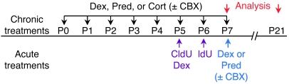
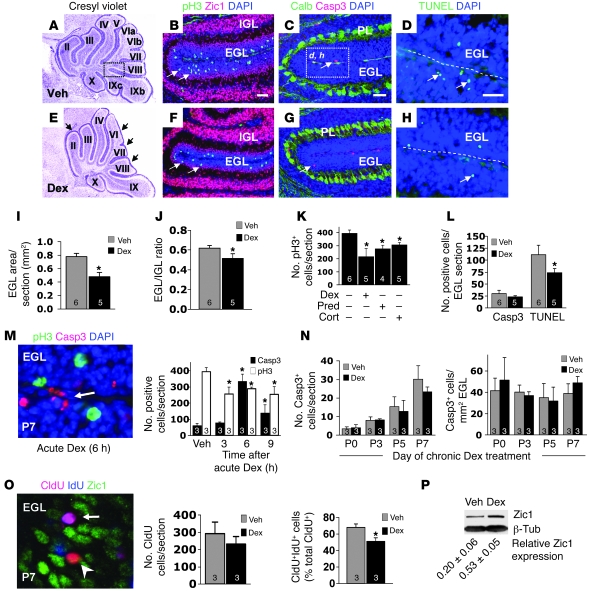
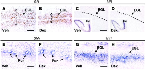
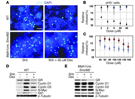
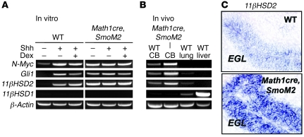
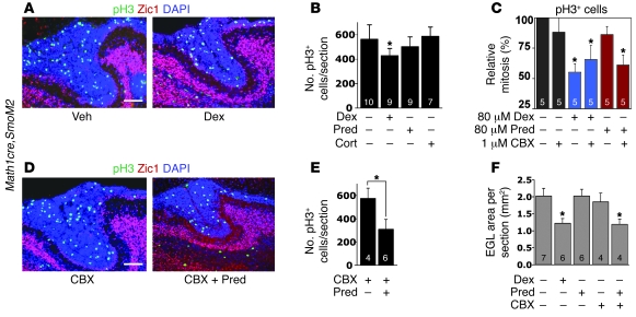
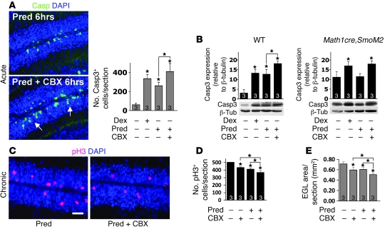
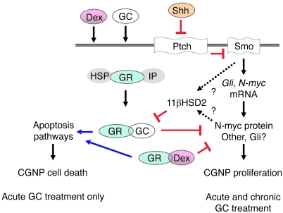
Glucocorticoids (GCs) are administered to human fetuses at risk of premature delivery and to infants with life-threatening respiratory and cardiac conditions. However, there are ongoing concerns about adverse effects of GC treatment on the developing human brain, although the precise molecular mechanisms underlying GC-induced brain injury are unclear. Here, we identified what we believe to be novel cross-antagonistic interactions of Sonic hedgehog (Shh) and GC signaling in proliferating mouse cerebellar granule neuron precursors (CGNPs). Chronic GC treatment (from P0 through P7) in mouse pups inhibited Shh-induced proliferation and upregulation of expression of N-myc, Gli1, and D-type cyclin protein in CGNPs. Conversely, acute GC treatment (on P7 only) caused transient apoptosis. Shh signaling antagonized these effects of GCs, in part by induction of 11beta-hydroxysteroid dehydrogenase type 2 (11betaHSD2). Importantly, 11betaHSD2 antagonized the effects of the GCs corticosterone, hydrocortisone, and prednisolone, but not the synthetic GC dexamethasone. Our findings indicate that Shh signaling is protective in the setting of GC-induced mouse neonatal brain injury. Furthermore, they led us to propose that 11betaHSD2-sensitive GCs (e.g., hydrocortisone) should be used in preference to dexamethasone in neonatal human infants because of the potential for reduced neurotoxicity.


Figures

Figure 1
Figure 1. Timing of chronic and acute treatment protocols.
Chronic GC administration from P0 to P7 is shown in black; acute GC treatment at P7 is shown in blue; administration of thymidine analogs CldU and IdU is shown in purple. Dex was given 2 h after CldU. Pups were analyzed to determine weight gain at P7 and P21 (see Table 1).
Figure 2
Figure 2. Dex treatment inhibits neonatal granule cell precursor proliferation.
(AH) P7 mouse pups were treated with vehicle (Veh; AD) or Dex (EH). (A and E) Chronic Dex treatment disrupted cerebellar foliation in lobes II–VII (arrows). Original magnification, ×20. The boxed region in A is shown at higher magnification in B, C, F, and G. (B and F) Immunocytochemistry for Zic1 and pH3 (arrows). (C and G) Immunocytochemistry for Casp3 (arrows). Dex did not change the mean total number of Calbindin-positive Purkinje cells (not shown). PL, Purkinje cell layer. The boxed region in C is shown at higher magnification in D and H. (D and H) TUNEL assay. Dotted lines denote the outer border of the EGL. (I and J) Mean EGL surface area per section (I) and proportional to the IGL (J). (K) pH3+ cells per sagittal section of mice treated with vehicle, Dex, Pred, or Cort. (L) Number of apoptotic cells in the EGL. (M) Number of Casp3+ and pH3+ cells after acute treatment. Original magnification, ×1,000. (N) Casp3+ cells during chronic treatment, both total number and per mm2 EGL. (O) Dex treatment 2 h after CldU injection (see Figure 1) did not affect the number of CldU+ cells, but significantly decreased the number of CldU+ldU+ cells. Original magnification, ×1,000. (P) Western blot analysis of protein lysates of whole cerebella. Zic1 protein levels, shown as estimated mean ± SD number of cells per sagittal section, significantly increased after Dex treatment (P < 0.01). Scale bars: 100 mm. For each group, n is shown within the corresponding bar. Asterisks denote significant differences versus respective vehicle groups; exact P values are shown in Results.
Figure 3
Figure 3. Chronic Dex treatment causes decreased GR expression but does not affect Shh and Gli1.
(A and B) CGNPs in the EGL have high expression of the GR. Immunocytochemistry for GR of the Dex-treated cerebellum indicates the decreased surface area of the EGL, as shown in Figure 2E. The width of the EGL is shown by dotted lines. (C and D) MR was not expressed in the cerebellum. Dotted lines denote the outer border of the EGL. Hc, hippocampus. Original magnification of insets, ×40. (EH) There was no change in Shh and Gli1 expression, as demonstrated by in situ hybridization of Shh-expressing (E and F) Purkinje (Pur) and Gli1-expressing (G and H) granule cells in vehicle- and Dex-treated mouse pups. Scale bars: 100 μm.
Figure 4
Figure 4. Cross-antagonistic Shh-GC signaling regulates CGNP proliferation in vitro.
(A) Immunocytochemistry for pH3 of CGNP cultures, generated from WT or Math1cre,SmoM2 animals, and treated with Shh alone or in combination with 40 μM Dex. (B) To determine the optimal dose to inhibit CGNP proliferation, cultures were exposed to different doses of Dex and Pred for 24 h. The number of pH3+ cells in the WT cultures significantly decreased with increasing Dex (black triangles) and were unaffected by Pred (black circles). The number of pH3+ cells in the Math1cre,SmoM2 cultures were not affected up to 40 μM Dex (blue circles). (C) At 60–160 μM Dex, the number of pH3+ cells in the Math1cre,SmoM2 cultures was dose-dependently affected (blue circles). Conversely, Math1cre,SmoM2 cultures were protected against Pred up to 120 μM (red squares). (D) Protein lysates were prepared from CGNPs treated with vehicle, Shh, or Shh plus 40 μM Dex for 24 h. Western blot analysis showed that Dex treatment decreased cyclin D1, N-myc, Gli1, and cyclin D2 protein expression. (E) Western blot analysis of Math1cre,SmoM2 CGNPs showed that cyclin D1 levels were greatly upregulated, independent of Shh administration, and were unchanged after 40 μM Dex treatment. Furthermore, cyclin D2, Gli1, and N-myc levels were unchanged. Samples were run on the same gel but were noncontiguous. Scale bar: 50 μm. Exact P values are shown in Results.
Figure 5
Figure 5. Shh signaling upregulates transcription of 11βHSD2 in vitro and in vivo.
(A) Whole RNA was isolated from WT and Math1cre,SmoM2 CGNP cultures treated with vehicle, Shh, or Shh plus 40 μM Dex for 24 h. Quantitative PCR analysis showed that Shh treatment potently induced the Shh targets N-myc and Gli1 as well as 11βHSD2 expression in WT CGNP cultures. Math1cre,SmoM2 CGNPs showed increased expression of N-myc, Gli1, and 11βHSD2 in both vehicle and Shh groups and was unchanged after Dex treatment. 11βHSD1 expression was below detectable levels. (B) In vivo, N-myc, Gli1, and 11βHSD2 levels were upregulated in the Math1cre,SmoM2 cerebellum (CB). (C) In situ hybridization showing specific expression of 11βHSD2 in the EGL of the P7 WT and Math1cre,SmoM2 cerebellum. Original magnification, ×400.
Figure 6
Figure 6. CGNPs in Math1cre,SmoM2 transgenic mice are protected against antiproliferative effects of GCs in an 11βHSD2-dependent manner in vitro and in vivo.
(A) Immunocytochemistry for pH3 and Zic1 in vehicle- and Dex-treated Math1cre,SmoM2 P7 mice. (B) Mean ± SD number of pH3+ cells per section in the whole EGL of Math1cre,SmoM2 mice. Animals were protected against Pred and Cort treatment, but only partly so against Dex. (C) To determine whether inhibition of 11βHSD2 by CBX could induce a decrease in CGNP proliferation by 11βHSD2-sensitive Pred in vitro, Math1cre,SmoM2 cultures were exposed to 80 μM Dex or Pred in the presence or absence of CBX. The number of pH3+ cells significantly decreased when CBX was added to 80 μM Pred. (D) Similar to A, with Math1cre,SmoM2 P7 mice treated with CBX alone or in combination with Pred. (E) Treatment of Pred in combination with CBX resulted in significantly decreased numbers of pH3+ cells. (F) Area measurements showed that the mean surface area of the EGL also significantly decreased in pups treated with Dex and with Pred plus CBX. Scale bars: 100 μm. For each group, n is shown within the corresponding bar. Asterisks denote significant differences versus respective vehicle groups (brackets denote comparisons other than with vehicle); exact P values are shown in Results.
Figure 7
Figure 7. Evidence that 11βHSD2 activity is protective against acute and chronic Pred-induced changes in the cerebellum.
(A and B) In WT mice, acute Pred and Dex treatment caused a significant increase in the number of Casp3+ cells (arrows), and a further significant increase was observed with Pred in combination with CBX. Original magnification, ×400. (B) Conversely, acutely Pred-treated Math1cre,SmoM2 mice were protected, unless CBX was also administered. Samples were run on the same gel but were noncontiguous. (C and D) With chronic treatment, Pred caused significant inhibition of proliferation; its effects were significantly exacerbated by CBX. Scale bar: 100 μm. (E) Comparable significant changes were found after measuring the EGL volume. For each group, n is shown within the corresponding bar. Asterisks denote significant differences versus respective vehicle groups (brackets denote comparisons other than with vehicle); exact P values are shown in Results.
Figure 8
Figure 8. Model for cross-antagonistic interactions of Hedgehog signaling and acute/chronic GC signaling in developing CGNPs.
Shh-Smo activation in proliferating CGNPs promotes cell cycle progression and induces 11βHSD2 expression, directly or indirectly, through regulation of its downstream activators N-Myc, Gli, and possibly others. In this respect, Shh-Smo signaling is protective against the acute proapoptotic and chronic antiproliferative effects of 11βHSD2-sensitive GCs such as Pred and Cort, but not Dex or β-methasone. HSP, heat shock protein; IP, immunophillin.

Comment in

Similar articles

Cited by

References

    1. Liggins G.C., Howie R.N. A controlled trial of antepartum glucocorticoid treatment for prevention of the respiratory distress syndrome in premature infants. Pediatrics. 1972;50:515–525. - PubMed
    1. Dalziel S.R., et al. Antenatal exposure to betamethasone: psychological functioning and health related quality of life 31 years after inclusion in randomised controlled trial. BMJ. 2005;331:665. - PMC - PubMed
    1. Murphy B.P., et al. Impaired cerebral cortical gray matter growth after treatment with dexamethasone for neonatal chronic lung disease. Pediatrics. 2001;107:217–221. doi: 10.1542/peds.107.2.217. - DOI - PubMed
    1. Baud O. Postnatal steroid treatment and brain development. Arch. Dis. Child. Fetal Neonatal Ed. 2004;89:F96–F100. doi: 10.1136/adc.2003.028696. - DOI - PMC - PubMed
    1. Bodensteiner J.B., Johnsen S.D. Cerebellar injury in the extremely premature infant: newly recognized but relatively common outcome. J. Child Neurol. 2005;20:139–142. doi: 10.1177/08830738050200021101. - DOI - PubMed

Publication types