Review
. 2009 Feb;10(2):144-51.
doi: 10.1038/embor.2008.256. Epub 2009 Jan 23.

Retroviral integrase superfamily: the structural perspective

Affiliations
Review

Retroviral integrase superfamily: the structural perspective

Marcin Nowotny. EMBO Rep. 2009 Feb.

Abstract

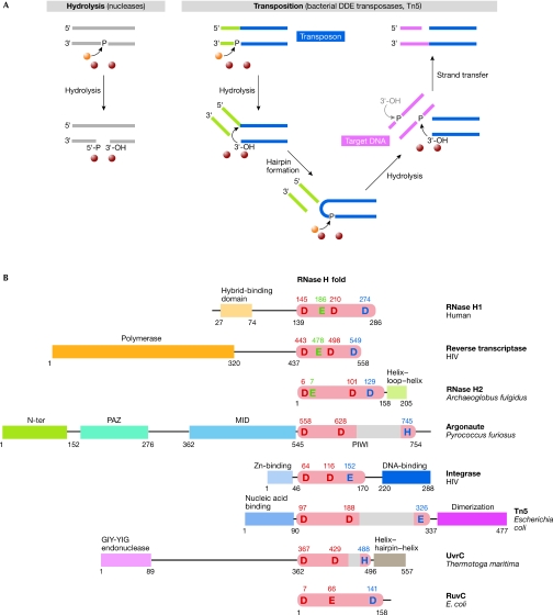
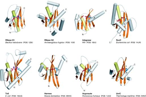
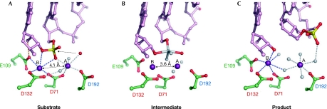
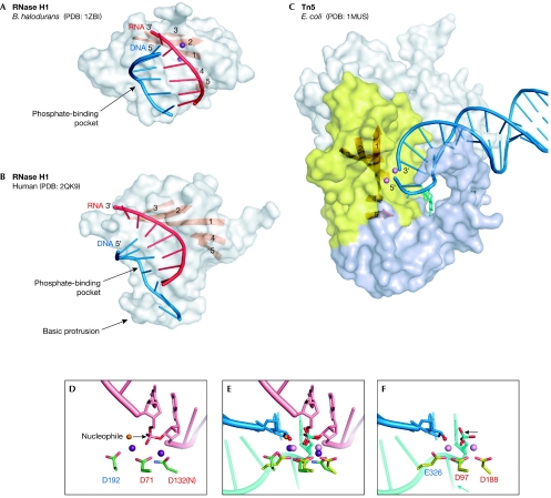
The retroviral integrase superfamily (RISF) comprises numerous important nucleic acid-processing enzymes, including transposases, integrases and various nucleases. These enzymes are involved in a wide range of processes such as transposition, replication and repair of DNA, homologous recombination, and RNA-mediated gene silencing. Two out of the four enzymes that are encoded by the human immunodeficiency virus--RNase H1 and integrase--are members of this superfamily. RISF enzymes act on various substrates, and yet show remarkable mechanistic and structural similarities. All share a common fold of the catalytic core and the active site, which is composed primarily of carboxylate residues. Here, I present RISF proteins from a structural perspective, describing the individual members and the common and divergent elements of their structures, as well as the mechanistic insights gained from the structures of RNase H1 enzyme complexes with RNA/DNA hybrids.


Figures

Figure 1
Figure 1
Examples of reactions catalysed by members of the retroviral integrase superfamily and domain representation of selected members. (A) Schematic representation of two reactions catalysed by the RISF: nucleic-acid hydrolysis and a transposition reaction. The processing of only one end of the transposon is shown for simplicity. The attacking 3′-OH on the other end of the transposon is shown in grey. Metal ions and the water molecule are shown as purple and orange spheres, respectively. (B) RISF members. The insertions in the RNase H fold are shown as grey boxes. The functions or names of other domains are given and the positions of the active-site residues are indicated. The red residues are the two spatially conserved carboxylates, and the blue residue is the last and more variant residue. RNase H-specific glutamates are shown in green. DDE, aspartate, aspartate, glutamate catalytic triad; HIV, human immunodeficiency virus; RISC, RNA-induced silencing complex; RISF, retroviral integrase superfamily; Tn5, transposon 5; Zn, zinc.
Figure 2
Figure 2
Catalytic core structures. The central β-sheet (with numbered strands) and the conserved α-helix are shown in orange and yellow, respectively. More divergent parts of the fold are shown in grey. The active-site residues are shown in ball-and-stick representation and are colour coded as described in Fig 1B. The two metal ions observed in the Tn5 and RNase H1 structures are shown in purple. The sites of insertions into the RNase H fold are shown as dashed lines. The direction of the last helix is indicated with an arrow. HIV, human immunodeficiency virus; PDB, Protein Data Bank code; Tn5, transposon 5.
Figure 3
Figure 3
The reaction catalysed by RNase H1 enzymes. (A) Substrate or pre-reactive state. The RNA is shown in pink ball-and-stick representation with the scissile phosphate shown in red and yellow, the attacking nucleophile is shown as a red sphere and the two Mg2+ ions as purple spheres. The contact between Glu (E)109 and the 2′-OH is shown as an orange line. The numbers of the active-site carboxylates are given and are colour-coded as described in Figs 1B and 2. (B) The transition state model based on a structure with a non-phosphorylated nick at the active site. The observed oxygen atoms are shown in red and the modelled transition state atoms are shown in grey. The dashed line indicates the bond that will be broken in the reaction. Note that the distance between the metal ions is shorter than in (A). (C) Product complex. After the reaction is complete, and the 5′-phosphate and 3′-OH are generated, the phosphate group is displaced from the active site. The representations are based on the structures of Bacillus halodurans RNase H1 enzyme in complex with RNA/DNA hybrids as follows: (A) partly inactive D192N mutant, Protein Data Bank (PDB) code ; (B) D192N mutant, PDB ; and (C) E188A mutant, PDB .
Figure 4
Figure 4
Nucleic-acid complexes of Bacillus halodurans and human RNase H1 enzymes, and Escherichia coli Tn5 transposase. (A) B. halodurans RNase H1. The protein is shown in surface representation with strands of the central β-sheet shown as an orange ribbon. The portion of the nucleic acid that interacts with the protein is shown in cartoon representation with the RNA shown in red and the DNA in blue. The two metal ions at the active site are shown as purple spheres. (B) Human RNase H1. (C) Tn5 transposase in complex with a resolved DNA hairpin (blue) corresponding to the transposon end. Only one subunit of the dimer is shown. The insertion in the RNase H fold is shown in light blue. The nucleotide that is flipped out to form the DNA hairpin and is stabilized by the interactions with the insertion domain is shown in cyan stick representation. The amino-terminal domain that interacts with the nucleic acids and the carboxy-terminal dimerization domain are shown in grey. Two Mn2+ ions are shown as pink spheres. (D) Close-up view of the active site of B. halodurans RNase H1. Active-site carboxylates are shown in green and are labelled using the same colour coding as in Figs 1B and 2. The RNA strand is shown in pink with the two nucleotides joined by the scissile phosphate shown in stick representation. Two Mg2+ ions are shown as purple spheres. The attacking nucleophile is shown as an orange sphere and the direction of the attack is indicated with an arrow. (E) Superposition of the active sites of RNase H1 anf Tn5 transposase (coloured as in (D) and (F)) based on the positions of C-α atoms of the active-site carboxylates. Note that although they were not included in the superposition, the phosphate groups, metal ions and attacking nucleophiles (water in B. halodurans RNase H1 and 3′-OH in E.coli Tn5) occupy similar positions. After the non-transferred strand (cyan) is removed from the Tn5 active site, this superposition might represent the configuration of the transposon end (3′-OH group) attacking the target DNA mimicked by the RNA strand (pink) from the B. halodurans RNase H1 structure. (F) Active site of Tn5 transposase. The DNA is shown in blue (transferred strand) and cyan (non-transferred strand). The Mn2+ ions are shown as pink spheres. The 3′-OH group is indicated with a red sphere. The direction of the postulated last nucleophilic attack by a water molecule to generate this 3′-OH is indicated with an arrow. The flipped-out base is indicated with a cyan arrow. PDB, Protein Data Bank code; Tn5, transposon 5.
None
Marcin Nowotny

References

    1. Ariyoshi M, Vassylyev DG, Iwasaki H, Nakamura H, Shinagawa H, Morikawa K (1994) Atomic structure of the RuvC resolvase: a Holliday junction-specific endonuclease from E. coli. Cell 78: 1063–1072 - PubMed
    1. Bennett RJ, Dunderdale HJ, West SC (1993) Resolution of Holliday junctions by RuvC resolvase: cleavage specificity and DNA distortion. Cell 74: 1021–1031 - PubMed
    1. Cerritelli SM, Frolova EG, Feng C, Grinberg A, Love PE, Crouch RJ (2003) Failure to produce mitochondrial DNA results in embryonic lethality in Rnaseh1 null mice. Mol Cell 11: 807–815 - PubMed
    1. Ceschini S, Keeley A, McAlister MS, Oram M, Phelan J, Pearl LH, Tsaneva IR, Barrett TE (2001) Crystal structure of the fission yeast mitochondrial Holliday junction resolvase Ydc2. EMBO J 20: 6601–6611 - PMC - PubMed
    1. Chapados BR, Chai Q, Hosfield DJ, Qiu J, Shen B, Tainer JA (2001) Structural biochemistry of a type 2 RNase H: RNA primer recognition and removal during DNA replication. J Mol Biol 307: 541–556 - PubMed

Publication types

MeSH terms