Review
. 2009 Feb;9(2):108-22.
doi: 10.1038/nrc2544.

A tense situation: forcing tumour progression

Affiliations
Review

A tense situation: forcing tumour progression

Darci T Butcher et al. Nat Rev Cancer. 2009 Feb.

Abstract

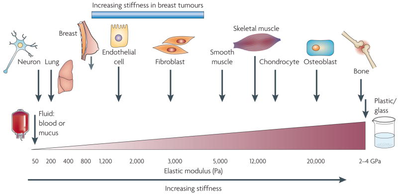
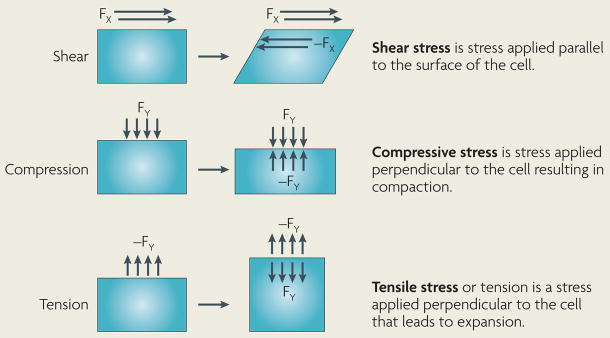
Cells within tissues are continuously exposed to physical forces including hydrostatic pressure, shear stress, and compression and tension forces. Cells dynamically adapt to force by modifying their behaviour and remodelling their microenvironment. They also sense these forces through mechanoreceptors and respond by exerting reciprocal actomyosin- and cytoskeletal-dependent cell-generated force by a process termed 'mechanoreciprocity'. Loss of mechanoreciprocity has been shown to promote the progression of disease, including cancer. Moreover, the mechanical properties of a tissue contribute to disease progression, compromise treatment and might also alter cancer risk. Thus, the changing force that cells experience needs to be considered when trying to understand the complex nature of tumorigenesis.


Figures

Figure 1
Figure 1. Cells are tuned to the materials properties of their matrix
All cells, including those in traditionally mechanically static tissues, such as the breast or the brain, are exposed to isometric force or tension that is generated locally at the nanoscale level by cell–cell or cell–extracellular matrix interactions and that influences cell function through actomyosin contractility and actin dynamics. Moreover, each cell type is specifically tuned to the specific tissue in which it resides. The brain, for instance, is infinitely softer than bone tissue. Consequently, neural cell growth, survival and differentiation are favoured by a highly compliant matrix. By contrast, osteoblast differentiation and survival occurs optimally on stiffer extracellular matrices with material properties more similar to newly formed bone. Normal mammary epithelial cell growth, survival, differentiation and morphogenesis are optimally supported by interaction with a soft matrix. Following transformation, however, breast tissue becomes progressively stiffer and tumour cells become significantly more contractile and hyper-responsive to matrix compliance cues. Normalizing the tensional homeostasis of tumour cells, however, can revert them towards a non-malignant phenotype, thereby illustrating the functional link between matrix materials properties, cellular tension and normal tissue behaviour. Importantly, however, although breast tumours are much stiffer than the normal breast, the materials properties of a breast tumour remain significantly softer than those of muscle or bone, emphasizing the critical association between tissue phenotype and matrix rigidity.
Figure 2
Figure 2. Mechanotransduction and focal adhesion maturation
a | The majority of integrins exist at the plasma membrane in a resting, inactive state in which they can be activated by inside–out or outside–in cues. With regard to outside–in activation, when cells encounter a mechanically rigid matrix or are exposed to an exogenous force integrins become activated, which favours integrin oligomerization or clustering, talin 1 and p130Cas protein unfolding, vinculin–talin association, and Src and focal adhesion kinase (FAK) stimulation of RhoGTPase-dependent actomyosin contractility and actin remodelling. Focal adhesions mature with the recruitment of a repertoire of adhesion plaque proteins, including α-actinin to facilitate actin association, and adaptor proteins such as paxillin, which foster interactions between multiple signalling complexes to promote growth, migration and differentiation. b | Normal cells tune their contractility in response to matrix stiffness cues, but tumours exhibit altered tensional homeostasis. Cells exert actomyosin contractility and cytoskeleton-dependent force in response to matrix stiffness cues. These forces can be measured using traction force microscopy. Thus, non-malignant human mammary epithelial cells spread more and exert more force on a stiff matrix than on a soft matrix. c | By comparison, breast tumour cells (T4) are highly contractile and spread considerably more than their non-malignant counterparts (S1) in response to the same compliant matrix. Importantly, inhibiting RhoGTPase signalling in tumour cells, by expressing a dominant-negative N19Rho or treating tumours with an inhibitor of Rho-associated, coiled-coil-containing protein kinase (ROCK; Y-27632) or myosin 2 (blebbistatin), reduces tumour cell contractility and spreading to levels exhibited by non-malignant breast epithelial cells. These data illustrate the importance of Rho signalling and actomyosin contractility in cell force generation and show how transformation alters cell force sensing. The traction map is shown in pseudocolour indicating regions of low (grey) and high (purple) forces in dynes per cm2. ECM, extracellular matrix; SFK, Src family kinase. Reproduced, with permission, from REF. © (2005) Elsevier Inc.
Figure 3
Figure 3. The normal mammary gland as a mechanically active tissue
a | The developing breast is subjected to a number of forces that facilitate its normal function. During lactation, for instance, the normal breast experiences compressive stress on the luminal epithelial cells and the basement membrane owing to the accumulation of milk and alveolar distension. Upon sucking and oxytocin stimulation, epithelial cells encounter inward tensile stress as the myoepithelium contracts to force the milk out of the alveolar sacs. In the absence of this stimulus, milk will accumulate within the acinus and eventually exert an outward projecting compressive force on the surrounding epithelium. This compressive force is countered by a compensatory inward projecting resistance force and the combination of these two forces eventually compromises the integrity of the tight junctions between alveolar cells. Chronic exposure to these forces and perturbed tissue integrin sensitize the gland to apoptotic cues so that the gland undergoes involution accompanied by extensive remodelling of the epithelium and the cellular and extracellular components of the stroma. b | Transformation (blue cells) resulting from the accumulation of genetic and epigenetic alterations in the epithelium along with an altered stromal matrix leads to unchecked proliferation and enhanced survival of luminal epithelial cells within the ductal tree, which compromises normal ductal architecture. With prolonged growth and abnormal survival, the abnormal pre-neoplastic luminal mammary epithelial cells eventually expand to fill the breast ducts. The expanding luminal epithelial mass exerts outward projecting compression forces of increasing magnitude on the basement membrane and adjacent myoepithelium. These forces are countered by an inward projecting resistance force. Importantly, the pre-neoplastic lesion secretes a plethora of soluble factors that stimulate immune cell infiltration and activation of resident fibroblasts to induce a desomoplastic response in the breast stroma. The desmoplastic stroma, which is characterized by dramatic changes in the composition, post-translational modifications and topology of the extracellular matrix (ECM), stiffens over time. This rigid parenchyma exerts a progressively greater inward projecting resistance force on the expanding pre-neoplastic duct. Over time, the number of myoepithelial cells surrounding the pre-neoplastic mass decreases and the basement membrane thins, probably owing to increased matrix metalloproteinase (MMP) activity, decreased protein deposition and compromised assembly (adapted from REF. 128). In parallel, there is a build-up of interstitial fluid pressure contributed by a leaky vasculature and compromised lymphatic drainage. In response to their genetic modifications and the altered materials properties of the matrix, the pre-neoplastic luminal epithelial cells exhibit modified tensional homeostasis and respond to the combination of forces and stromal cues to invade the breast parenchyma. Some resident fibroblasts transdifferentiate into myofibroblasts and facilitate tumour migration and invasion by promoting the assembly of linearized collagen fibrils surrounding the distended pre-neoplastic epithelial ducts.
Figure 4
Figure 4. Matrix stiffness modulates cellular morphology and epidermal growth factor (EGF)-dependent growth
Phase contrast microscopy and confocal immunofluorescence images of non-malignant immortalized human mammary epithelial cell (HMEC; MCF10A) colonies interacting with a three-dimensional reconstituted basement membrane (BM)-laminated polyacrylamide gel of increasing stiffness (150–5,000 Pa) showing colony morphogenesis after 20 days of culture. On compliant gels with materials properties similar to that measured in the normal murine mammary gland (150 Pa) non-malignant MECs proliferate for 6–12 days to eventually form growth-arrested, polarized acini analogous to the terminal ductal lobular units observed at the end buds of the differentiated breast. These structures have intact adherens junctions and insoluble cell–cell localized β-catenin before (main images) and after (inset a) Triton extraction, and polarity, as shown by the basal localization of (α6) β4 integrin, the apical–lateral localization of cortical actin (Phalloidin), and the assembly of an endogenous laminin 5 basement membrane. Incremental stiffening of the basement membrane gel progressively compromises tissue morphogenesis and alters EGF-dependent growth of these cells. Thus, colony size progressively increases with matrix stiffening, lumen formation is compromised, cell–cell junctions are disrupted, as revealed by loss of cell–cell-associated β-catenin (inset b), and tissue polarity is inhibited, as indicated by disorganized (α6) β4 integrin localization and loss of the endogenous laminin 5 basement membrane. Interestingly, actin stress fibres were not observed in the structures until the stiffness of the matrix reached 5,000 Pa, as has been observed in murine breast tumours in vivo. The arrows indicate loss of the endogenous basement membrane and disruption of basal polarity. Reproduced, with permission, from REF. © (2005) Elsevier Inc.
Figure 5
Figure 5. Imaging elastography of a breast tumour
Tissue imaging elastography is a spatial ‘visual’ qualitative measurement of the stiffness of a tissue that is generated by extrapolating tissue viscoelastic characteristics from ultrasound wave reflection in real-time. Photographs of sonoelastography images compare an elastogram image (a) with a B mode ultrasound scan (b) of a breast tumour. Ultrasound imaging elastography, as shown here, is an in situ mechanical imaging method that could improve the sensitivity and the specificity of breast cancer detection and may be a useful tool to advance our understanding of the link between mammographic density and the matrix materials properties of the breast. Image courtesy of A. Thomas & T. Fischer, Charité, Berlin, Germany.
None

References

    1. Gieni RS, Hendzel MJ. Mechanotransduction from the ECM to the genome: are the pieces now in place? J Cell Biochem. 2008;104:1964–1987. - PubMed
    1. Engler AJ, et al. Myotubes differentiate optimally on substrates with tissue-like stiffness: pathological implications for soft or stiff microenvironments. J Cell Biol. 2004;166:877–887. - PMC - PubMed
    1. Engler AJ, Sen S, Sweeney HL, Discher DE. Matrix elasticity directs stem cell lineage specification. Cell. 2006;126:677–689. Contractile myocytes were used to demonstrate that cells sense their mechanical environment. Myotubes form independently of matrix stiffness but myosin–actin striations emerge only on gels with stiffness typical of normal muscle, and not on matrices that are softer or stiffer. - PubMed
    1. Georges PC, Miller WJ, Meaney DF, Sawyer ES, Janmey PA. Matrices with compliance comparable to that of brain tissue select neuronal over glial growth in mixed cortical cultures. Biophys J. 2006;90:3012–3018. - PMC - PubMed
    1. McBeath R, Pirone DM, Nelson CM, Bhadriraju K, Chen CS. Cell shape, cytoskeletal tension, and rhoa regulate stem cell lineage commitment. Dev Cell. 2004;6:483–495. - PubMed

Publication types