. 2009 Jan;5(1):e1000270.
doi: 10.1371/journal.ppat.1000270. Epub 2009 Jan 23.

Rab11A-controlled assembly of the inner membrane complex is required for completion of apicomplexan cytokinesis

Affiliations

Rab11A-controlled assembly of the inner membrane complex is required for completion of apicomplexan cytokinesis

Carolina Agop-Nersesian et al. PLoS Pathog. 2009 Jan.

Abstract

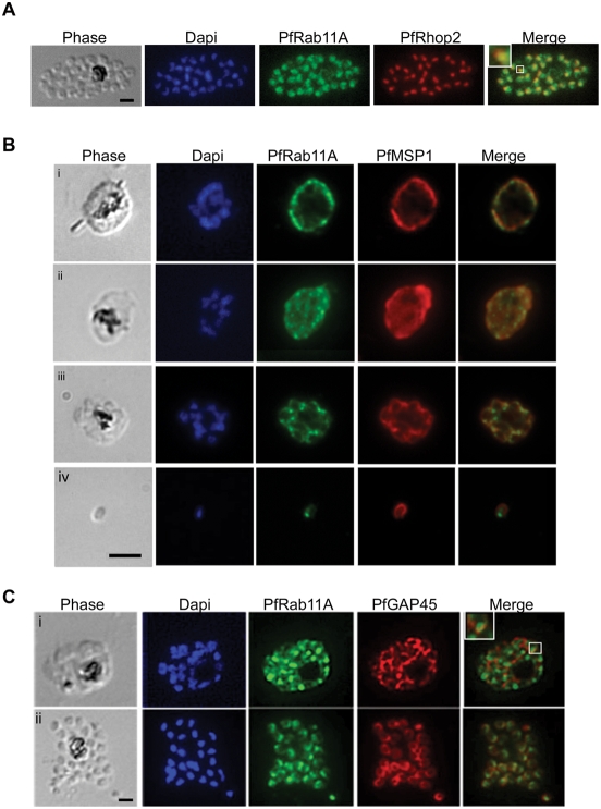
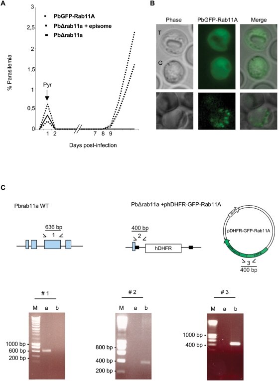
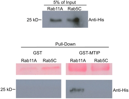
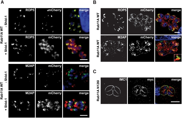
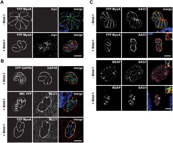
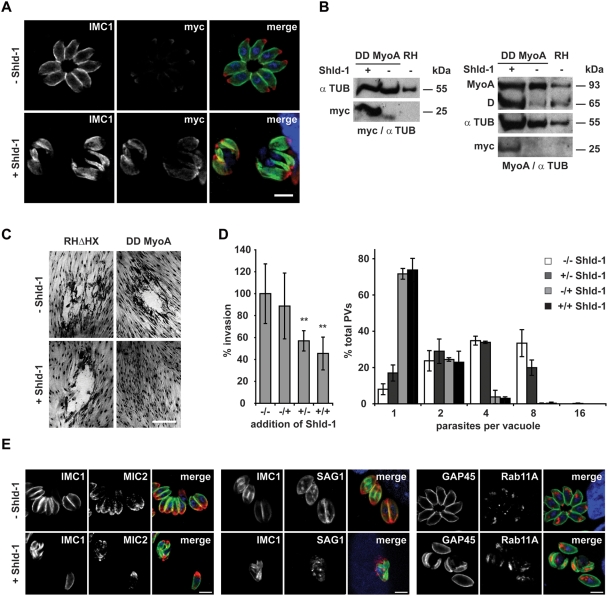
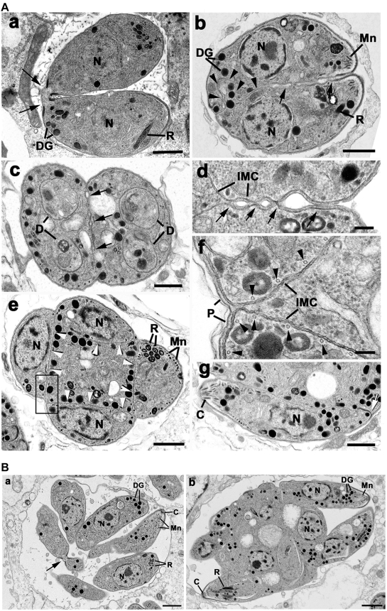
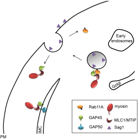
The final step during cell division is the separation of daughter cells, a process that requires the coordinated delivery and assembly of new membrane to the cleavage furrow. While most eukaryotic cells replicate by binary fission, replication of apicomplexan parasites involves the assembly of daughters (merozoites/tachyzoites) within the mother cell, using the so-called Inner Membrane Complex (IMC) as a scaffold. After de novo synthesis of the IMC and biogenesis or segregation of new organelles, daughters bud out of the mother cell to invade new host cells. Here, we demonstrate that the final step in parasite cell division involves delivery of new plasma membrane to the daughter cells, in a process requiring functional Rab11A. Importantly, Rab11A can be found in association with Myosin-Tail-Interacting-Protein (MTIP), also known as Myosin Light Chain 1 (MLC1), a member of a 4-protein motor complex called the glideosome that is known to be crucial for parasite invasion of host cells. Ablation of Rab11A function results in daughter parasites having an incompletely formed IMC that leads to a block at a late stage of cell division. A similar defect is observed upon inducible expression of a myosin A tail-only mutant. We propose a model where Rab11A-mediated vesicular traffic driven by an MTIP-Myosin motor is necessary for IMC maturation and to deliver new plasma membrane to daughter cells in order to complete cell division.


Conflict of interest statement

The authors have declared that no competing interests exist.

Figures

Figure 1
Figure 1. PfRab11A has a dynamic localisation.
(A–C) Throughout the erythrocytic stage of parasite development the localisation of PfRab11A changes. (A) Rhoptry localisation was confirmed in mature schizonts by immuno-staining infected blood smears with anti-PfRab11A and co-localisation (enlargement shown boxed in merge) demonstrated with anti-PfRhopH2 (rhoptry specific marker) antibodies. The bright-field image shows a mature schizont with the nuclei stained with Dapi. (B) PfRab11A co-localises partially with PfMSP1 (plasma membrane specific marker) in trophozoites (i, ii) and schizonts (iii). The merged images show co-localisation of the two proteins, giving single dots. In merozoites (iv), the localisation of PfMSP1 and PfRab11A is now distinct with PfRab11A having an apical localisation slightly under the plasma membrane decorated by PfMSP1. (C) The first two panels show the bright-field (phase) and Dapi, respectively. The last panel in all images shows an overlay. In young (i) and segmented (ii) schizonts, PfRab11A partially co-localises with PfGAP45 (Glidosome Associated Protein 45 used as an Inner Membrane Complex specific marker). In all cases, the black scale bar in the first panel of each set of images represents 5 µm.
Figure 2
Figure 2. The P. berghei rab11a gene is essential for growth in red blood cells.
(A) Growth comparison of transgenic parasites. To disrupt the rab11a locus, P. berghei merozoites were electroporated with the HindIII/EcoRI-linearized replacement plasmid containing 5′- and 3′-untranslated regions (black boxes in C) of rab11a and the human dhfr selectable marker (hDHRF); electroporated merozoites were subsequently injected into young mice. Drug selection of transfectants was by pyrimethamine (Pyr) addition to the drinking water 24 h post-transfection. No PbDHΔ11a disrupted parasites were recovered, only transfection of the linearized plasmid together with phDHFR-GFPRab11A vector (episome) generated transgenic parasites. The growth of rescued parasites harbouring the PbΔrab11a+episome is slower compared to parasites harbouring just episomal PbGFP-Rab11A. (B) PbRab11A localisation in trophozoites (T) and gametocytes (G) appears cytoplasmic, whereas in schizonts (lower panel) it appears more vesicular rhoptry-like. (C). Replacement-specific PCR analysis: A wild type specific PCR using primer combinations indicated by (1) was performed to confirm the disruption of the endogenous gene (exons shown in blue) in the clonal rescued parasite population. Confirmation of integration into the Pbrab11a locus and the presence of the episome were achieved by specific primer combinations (2 and 3), which detect the recombinant locus and the episome in transgenic parasites. The products run on 1% (wt/vol) agarose gel were stained with ethidium bromide. The molecular sizes are indicated in base pair (bp). The expected size of the respective PCR product is indicated in the diagram. M: marker; a and b: DNA isolated from wt and PbΔrab11a (+episome) parasites, respectively.
Figure 3
Figure 3. PfRab11A associates with PfMTIP in vitro.
The purified recombinant proteins PfRab11A-His and PfRab5C-His (top panel) were mixed with GST or GST–MTIP (bottom panel). Anti-His antibodies (Santa Cruz) were used to detect pulled-down His-tagged Rab protein and only PfRab11A associates with PfMTIP. The upper panel of the pull down correspond to Ponceau S stain of the membrane to demonstrate loadings of GST and GST-MTIP, respectively.
Figure 4
Figure 4. Overexpression of the wild type and dominant-negative Rab11A in T. gondii.
(A) Regulation of ddFKBP-mCherry-Rab11Awt expression in stable transfected parasites. Parasites were inoculated on HFF cells in presence and absence of 1 µM Shld-1 for 16 hours and probed with the indicated antibodies. In the absence of Shld-1, weak background fluorescence can be detected that co-localises with the rhoptry marker Rop5. Overexpression (+Shld-1) of Rab11Awt results in partial accumulation in an endosome-associated compartment as indicated by partial co-localisation with proM2AP . Scale bar: 5 µm. (B) Confocal microscopy of the same parasite strain as in (A). High-resolution microscopy reveals that overexpression of Rab11A results in association with a “vesicular network” to which the rhoptries appear to align. Again a partial co-localisation with proM2AP is evident. Scale bar: 5 µm. (C) Overexpression of dominant negative Rab11A(N126I) N-terminally tagged with ddFKBPmyc results in a different, vesicular localisation that accumulates at the IMC as indicated by partial co-localisation with antibody against IMC1 . Note that no accumulation is evident at the rhoptries (see also Figure S3). Scale bar is 5 µm. The green and red colours in the merged images correspond to the left and middle panels, respectively.
Figure 5
Figure 5. Expression of Rab11A(N126I) results in a specific effect on the formation of the glideosome.
Parasites expressing ddFKBPRab11A(N126I) were inoculated on host cells (HFF). After invasion, intracellular parasites were treated with or without 1 µM Shld-1 for 16 hours, before samples were fixed and stained with the indicated antibodies. (A) Parasites co-expressing YFP-MyoA and ddFKBPRab11A(N126I) were analysed. Note that tagged Rab11A(N126I) (as detected with myc-antibodies) specifically accumulates at regions where less YFP-MyoA is detectable within the IMC (arrow). (B). Analysis of further components of the glideosome/IMC in parasites expressing ddFKBPRab11A(N126I). Top: Parasites co-expressing YFP-GAP50 and ddFKBPRab11(N126I). Note that GAP50 can be identified at the IMC of first- and premature second-generation daughter parasites. In contrast, GAP45 (as detected with GAP45 antibodies) can mainly be identified at the IMC of the original mother parasite. Only a faint staining of the first-generation daughter cells is detectable. Middle: Same experiment with parasites co-expressing IMC1-YFP and ddFKBPRab11A(N126I). The localisation of MLC1 is restricted to the first-generation mother cell, whereas IMC1 can be detected at the IMC of first- and second-generation daughter cells. Bottom: Same parasites as in A). Both, MyoA and MLC1 show mainly staining of the mother parasite. (C) Expression of Rab11A(N126I) results in intracellular accumulation of SAG1. Upper panels: Parasites co-expressing YFP-MyoA and ddFKBPRab11A(N126I) were stained using monoclonal SAG1 antibodies. Whereas in the absence of Shld-1 parasite replication was evident with SAG1 exclusively detected at the surface, stabilisation of Rab11A(N126I) resulted in intracellular localisation of SAG1. Note that even plasmamembrane-associated SAG1 shows a rather patchy pattern. Lower panels: Parasites expressing ddFKBPRab11A(N126I) have been grown under the same conditions and a co-localisation with antibodies against proM2AP and SAG1 has been performed. Internalised SAG1 partially co-localises with proM2AP positive endosomes (arrows). In the upper corners, an enlarged area of the boxed area is shown. Scale bars: 5 µm. The green and red colours in the merged images correspond to the left and middle panels, respectively.
Figure 6
Figure 6. Inducible expression of a dominant negative MyoA results in a similar phenotype as observed for Rab11A(N126I).
(A). Stable transfection of wild type parasites with the construct p5RT70ddFKBPMyoAtail allows specific and inducible regulation of ddFKBPMyoAtail in dependence of Shld-1. Parasites were inoculated on HFF cells in the presence and absence of 1 µM Shld-1 and stained with the indicated antibodies. In the absence of Shld-1, weak background levels of ddFKBPMyoAtail can be detected close to the apical pole of the parasite. In the presence of Shld-1, high protein levels can be detected that co-localise with the IMC. Not that these parasites show a very abnormal IMC when compared to non-treated parasites. Scale bar: 5 µm. The green and red colours in the merged images correspond to the left and middle panels, respectively. (B) Immunoblot analysis of parasites stably transfected with p5RT70ddFKBPMyoAtail. Parasites were incubated for 5 hours in the presence or absence of Shld-1 before lysates were prepared. Left: Blot was simultaneously probed with myc- and alpha-Tubulin (as loading control) antibodies to analyze Shld-1 dependent regulation of ddFKBPMyoAtail. Right: Blot was simultaneously probed with antibodies against MyoA and alpha-Tubulin. Occasionally we detected a degradation product (D) of endogenous MyoA at ∼65 kDa that appears to be more prominent when ddFKBPMyoAtail is stabilized. (C) Stabilization of ddFKBPMyoAtail is deleterious for the parasite. Stable transfected parasites were inoculated on HFF cells in the presence and absence of 1 µM Shld-1 for 6 days before formation of plaques was compared. In the presence of Shld-1, no growth of parasites was detected. Scale bar 20 µm. (D). Invasion and replication analysis of parasites expressing ddFKBPMyoAtail. (−/−) parasites without Shld-1 treatment; (−/+) parasites treated with Shld-1 after invasion; (+/−) parasites treated with Shld-1 before invasion and without Shld-1 after invasion; (+/+) parasites constantly kept under Shld-1 treatment. Left: For the invasion assay, the total number of parasitophorous vacuoles was determined. Mean values of 5 (+/− and −/+) and 6 (−/− and +/−) independent experiments ±s.d. are shown. Asterisks indicate significant difference in total invasion compared to parasite strain ddFKBPMyoAtail not treated with Shld-1 before and after invasion (P<0.01, two tailed Student's t-test). Right: For the replication assay the number of parasites per parasitophorous vacuole was determined. Mean values of three independent experiments are shown +/−s.d. (E) Immunofluorescence analysis of parasites stably transfected with p5RT70ddFKBPMyoAtail. Parasites were treated with Shld-1 after invasion of the host cell (+) or left untreated (−). 16 hours post invasion, parasites were fixed and analysed with the indicated antibodies. Scale bars: 5 µm. The green and red colours in the merged images correspond to the left and middle panels, respectively.
Figure 7
Figure 7. Transmission electron micrographs of parasites expressing ddFKBPRab11A(N126I) treated with or without Shld-1.
(A) Transmission electron micrographs of samples not treated with Shld-1 (a) and treated with Shld-1 for 24 hours (b–g). (a) Longitudinal section through a control sample showing two daughters formed by endodyogeny still connected by their posterior end (arrows). Bar is 1 µm. (b) Longitudinal section through a sample treated with Shld-1 illustrates two fully formed daughters incompletely separated along their lateral surface (arrows) and with an irregular IMC towards the posterior (arrowheads). Bar is 1 µm. (c) Cross section through two incompletely separate daughters (arrows) showing the initiation of a second round of endodyogeny with the formation of the two IMC of the second generation of daughters (D) within each first-generation daughter. Bar in 1 µm. (d) Enlargement of the lateral surface between two daughters at a similar stage to that in (b) in which vesicles appear to form between the IMC but do not fuse (arrows) to form the plasmalemma. IMC, inner membrane complex. Bar is 100 nm. (e) Cross section through a late stage in the second round of endodyogeny showing the four daughters located around the periphery of the organism each separated by an IMC (arrowheads) leave a central cytoplasmic mass. Bar is 1 µm. (f) Detail from an area similar to the enclosed showing the IMC to consist of two unit membranes with underlying microtubles (arrowheads). Note the interaction of the IMC with the plasmalemma (P) at the external surface to form the pellicle, but the absence of any vesicular formation associated with internal regions. Bar is 100 nm. (g) A section through the periphery of a similar stage to that in (e) showing a longitudinal section of a tachyzoite with normal organelles and thickening of the IMC at the posterior pore (arrow), but still connected along its lateral surface. Bar is 1 µm. (B) (a) Section through a control sample showing the multiple daughters formed by repeated cycles of endodyogeny arranged in a rosette. Note certain daughters still connected by the posterior end (arrow). C, conoid. Bar is 1 µm. (b) Section through a sample treated with Shld-1 for 36 hours showing a large cytoplasmic mass with numerous nuclei and the partial formation of the anterior end of certain tachyzoites. Bar is 1 µm. C, conoid; DG, dense granule; Mn, microneme; N, nucleus; R, rhoptry.
Figure 8
Figure 8. Model of Rab11A function during the late stage of parasite replication.
Rab11A interacts with components of the glideosome (MLC/MTIP, GAP45 and MyoA) and transports vesicles derived from the secretory pathway to the novel synthesized plasma membrane of the daughter cells. Upon fusion of the vesicle with the PM, the complex is disassembled. Whereas Rab11A is recycled for the next round of transport, components of the proto-glideosome are freed to assemble and interact with early components of the glideosome in the IMC (like GAP50).

References

    1. D'Avino PP, Savoian MS, Capalbo L, Glover DM. RacGAP50C is sufficient to signal cleavage furrow formation during cytokinesis. J Cell Sci. 2006;119:4402–4408. - PubMed
    1. D'Avino PP, Savoian MS, Glover DM. Cleavage furrow formation and ingression during animal cytokinesis: a microtubule legacy. J Cell Sci. 2005;118:1549–1558. - PubMed
    1. Jurgens G. Plant cytokinesis: fission by fusion. Trends Cell Biol. 2005;15:277–283. - PubMed
    1. Ferguson DJ, Sahoo N, Pinches RA, Bumstead JM, Tomley FM, et al. MORN1 has a conserved role in asexual and sexual development across the apicomplexa. Eukaryot Cell. 2008;7:698–711. - PMC - PubMed
    1. Striepen B, Jordan CN, Reiff S, van Dooren GG. Building the perfect parasite: cell division in apicomplexa. PLoS Pathog. 2007;3:e78. doi:101371/j doi:10.1371/journal.ppat.0030078. - PMC - PubMed

Publication types

MeSH terms

LinkOut - more resources