. 2009;4(1):e4255.
doi: 10.1371/journal.pone.0004255. Epub 2009 Jan 23.

Nuclear envelope remnants: fluid membranes enriched in sterols and polyphosphoinositides

Affiliations

Nuclear envelope remnants: fluid membranes enriched in sterols and polyphosphoinositides

Marie Garnier-Lhomme et al. PLoS One. 2009.

Abstract

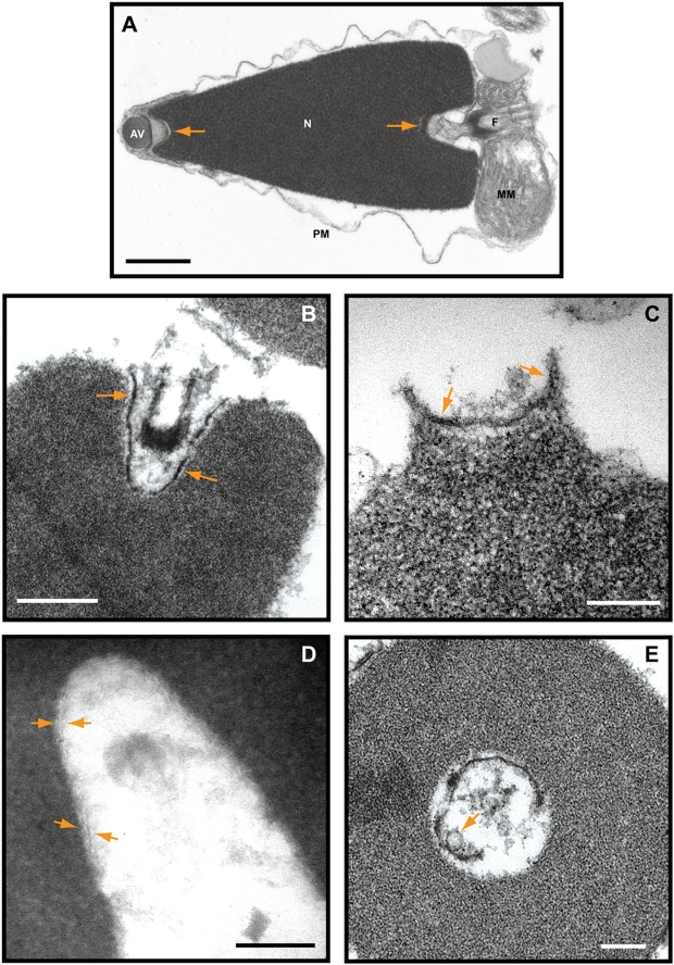
Background: The cytoplasm of eukaryotic cells is a highly dynamic compartment where membranes readily undergo fission and fusion to reorganize the cytoplasmic architecture, and to import, export and transport various cargos within the cell. The double membrane of the nuclear envelope that surrounds the nucleus, segregates the chromosomes from cytoplasm and regulates nucleocytoplasmic transport through pores. Many details of its formation are still unclear. At fertilization the sperm devoid of nuclear envelope pores enters the egg. Although most of the sperm nuclear envelope disassembles, remnants of the envelope at the acrosomal and centriolar fossae do not and are subsequently incorporated into the newly forming male pronuclear envelope. Remnants are conserved from annelid to mammalian sperm.

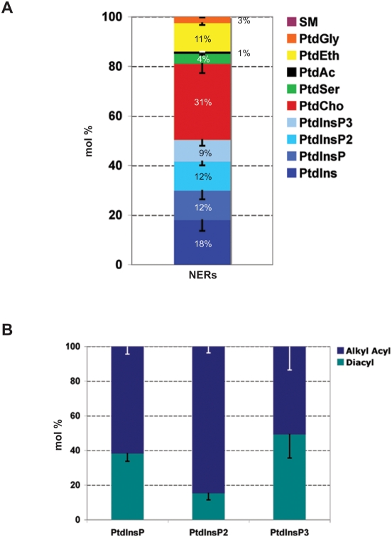
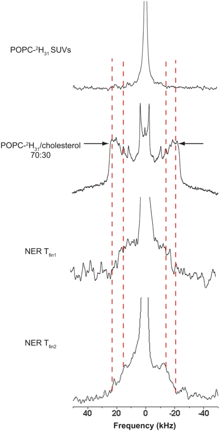
Methodology/principal findings: Using lipid mass spectrometry and a new application of deuterium solid-state NMR spectroscopy we have characterized the lipid composition and membrane dynamics of the sperm nuclear envelope remnants in isolated sperm nuclei.

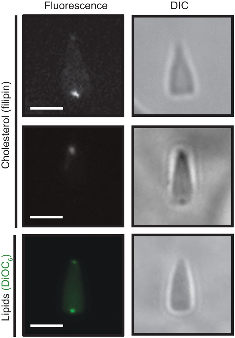
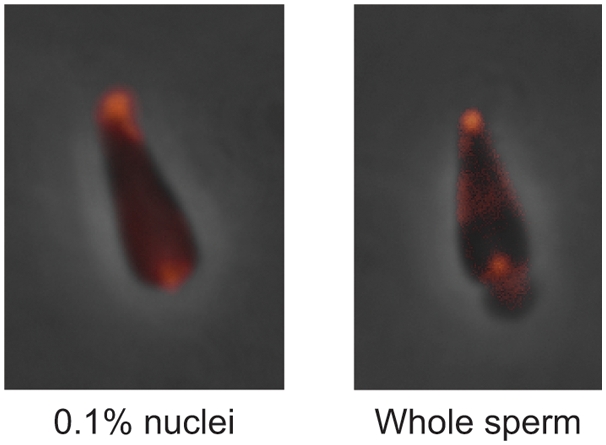
Conclusions/significance: We report nuclear envelope remnants are relatively fluid membranes rich in sterols, devoid of sphingomyelin, and highly enriched in polyphosphoinositides and polyunsaturated phospholipids. The localization of the polybasic effector domain of MARCKS illustrates the non-nuclear aspect of the polyphosphoinositides. Based on their atypical biophysical characteristics and phospholipid composition, we suggest a possible role for nuclear envelope remnants in membrane fusion leading to nuclear envelope assembly.


Conflict of interest statement

Competing Interests: The authors have declared that no competing interests exist.

Figures

Figure 1
Figure 1. Nuclear envelope remnants contain two membranes that line the acrosomal and centriolar fossae.
(A) P. lividus sperm cells were fixed in the presence of 1% (w/v) tannic acid. The plasma membrane (PM) and the mitochondrial membranes (MM) are shown. AV: acrosomal vesicle, N: nucleus, F: flagellum. The nuclear envelope is tightly apposed to the chromatin but cup-like structures with nuclear envelope remnants can be seen at the poles (arrows). (B and C) S. purpuratus 0.1% nuclei were incubated in egg cytoplasm supplemented with ATP-GS and fixed in the presence of 1% (w/v) tannic acid. Electron dense structures (arrows) are shown in the centriolar (B) and acrosomal fossae (C). The two bilayers appear to have variable amounts of electron dense material between them. (D) Cryosections of S. purpuratus 0.1% nuclei prefixed in 4% (v/v) formaldehyde for 3h on ice show two membranes in the centriolar fossa (arrows). (E) S. purpuratus 0.1% nuclei were incubated in egg cytoplasm in the presence of an ATP-generating system, fixed in 2.5% (v/v) glutaraldehyde in the presence of 1% (w/v) tannic acid and viewed by TEM. The glancing cross section of the centriolar fossa shows the nuclear envelope remnants and an egg membrane vesicle (arrow) associated with the nuclear envelope remnants. Bars are 500nm (A), 400nm (B) and 200nm (C, D and E). The data are representative of nuclei observed in at least 3 experiments on independent nuclei preparations.
Figure 2
Figure 2. Cholesterol and phospholipids both localize at the poles of the sperm nucleus.
The 0.1% nuclei were fixed in 4% (v/v) formaldehyde at 4°C and stained either with filipin to label cholesterol or DiOC6 to label nuclear envelope remnants lipids. Cholesterol labelling co-localizes with nuclear envelope remnants at both the acrosomal and centriolar fossae. DIC: differential interference contrast. Bars are 2.5 μm. Note that two panels of filipin staining are shown as in each case only one NER was in the confocal plane. Data are representative of at least 3 experiments performed on independent nuclei preparations.
Figure 3
Figure 3. Nuclear envelope remnants are enriched in polyphosphoinositides.
Lipid analysis of nuclear envelope remnants. (A) Lipids extracted from L. pictus demembranated sperm cells were separated by HPLC on a normal phase column and characterized by ESI-MS/MS using the precursor ion scans of sphingomyelin (SM), phosphatidylglycerol (PtdGly), phosphatidylethanolamine (PtdEth), phosphatidic acid (PtdAc), phosphatidylserine (PtdSer), phosphatidylcholine (PtdCho), phosphatidylinositol (PtdIns) or using the multiple ion scans of phosphatidylinositolphosphate (PtdInsP), phosphatidylinositolbisphosphate (PtdInsP2) and phosphatidylinositoltrisphosphate (PtdInsP3). Phospholipids were quantified using 12∶0/12∶0 (SM, PtdGly, PtdEth, PtdAc, PtdSer and PtdCho) or 16∶0/16∶0 (PtdIns, PtdInsP, PtdInsP2 and PtdInsP3) internal standards. Data expressed as mean±SEM (n = 3). (B) Alkyl-acyl versus diacyl phosphoinositides species distribution in nuclear envelope remnants. Mole percentages of diacyl species (green) and alkyl-acyl species (blue) were quantified from the multiple ion scans for each phosphoinositide class: PtdInsP, PtdInsP2 and PtdInsP3. 38% of PtdInsP, 15% of PtdInsP2 and 49% of PtdInsP3 are diacyl species. The PtdInsP2 is predominantly alkyl-acyl phosphoinositide. Data expressed as mean±SEM (n = 3).
Figure 4
Figure 4. Poly-phosphoinositides in 0.1% nuclei and whole sperm are enriched in the acrosomal and centriolar fossae.
L. pictus 0.1% nuclei (left) and whole live sperm (right) were incubated with the Texas Red labelled MARCKS peptide and visualised by fluorescence microscopy. The punctate staining of the acrosomal and centriolar fossae is typical of the majority of nuclei observed in experiments on two independent sperm and 0.1% nuclei preparations.
Figure 5
Figure 5. Nuclear envelope remnants are relatively fluid membranes despite their enrichment in cholesterol.
The top solid-state deuterium NMR spectrum corresponds to POPC-2H31 small unilamellar vesicles (SUV) acquired at 10°C post equilibrium at 40°C for 22h. The second spectrum corresponds to POPC-2H31 MLVs containing 30 mol% cholesterol acquired at 10°C after equilibration at 40°C. The bottom two spectra are of 0.1% nuclei incubated with SUVs of deuterated lipid (POPC-2H31) for 30 min at 40°C. Labelled nuclear envelope remnants were equilibrated at 40°C for 22h and the 2H NMR spectrum was acquired post-equilibrium at 10°C for 4h (NER Tfin1) and 20h (NER Tfin2). The dashed lines show the plateau quadrupolar splitting enlargement of labelled nuclear envelope remnants post-equilibrium. Arrows show the plateau quadrupolar splittings used to calculate the order parameters. NMR spectra are representative of those obtained in a duplicate experiment.
Figure 6
Figure 6. Cholesterol removal from nuclear envelope remnants inhibits membrane fusion events of nuclear envelope formation.
(A) 0.1% L. pictus nuclei were treated with 10mM MβCD or untreated (control). Nuclei were filipin stained, normalized to an equal nuclei concentration and excited at 360nm. The fluorescence intensity of the emission peak at 479/480nm was measured, and data were normalized to the control value. Data shown are mean±SEM of four experiments conducted in duplicate. (B) 0.1% L. pictus nuclei were treated with 10mM MβCD (white) or untreated (black). Nuclear envelope precursor MVs were subsequently bound to nuclei (ATP), and in parallel reactions their fusion was triggered with GTP to complete envelope formation. At least 20 nuclei were scored for the presence of a fully-formed nuclear envelope in 3 independent experiments. Data shown are mean±SEM.

References

    1. Longo FJ, Anderson E. The fine structure of pronuclear development and fusion in the sea urchin, Arbacia punctulata. J Cell Biol. 1968;39:339–368. - PMC - PubMed
    1. Collas P, Poccia DL. Conserved binding recognition elements of sperm chromatin, sperm lipophilic structures and nuclear envelope precursor vesicles. Eur J Cell Biol. 1996;71:22–32. - PubMed
    1. Colwin AL, Colwin LH. Changes in the spermatozoon during fertilization in Hydroides hexagonus (Annelida). II. Incorporation with the egg. J Biophys Biochem Cytol. 1961;10:255–274. - PMC - PubMed
    1. Collas P, Poccia D. Lipophilic organizing structures of sperm nuclei target membrane vesicle binding and are incorporated into the nuclear envelope. Dev Biol. 1995;169:123–135. - PubMed
    1. Cameron LA, Poccia DL. In vitro development of the sea urchin male pronucleus. Dev Biol. 1994;162:568–578. - PubMed

Publication types

MeSH terms

Substances