

Lactational coumestrol exposure increases ovarian apoptosis in adult rats
- PMID: 19165469
- PMCID: PMC2695544
- DOI: 10.1007/s00204-008-0400-0
Lactational coumestrol exposure increases ovarian apoptosis in adult rats
Abstract
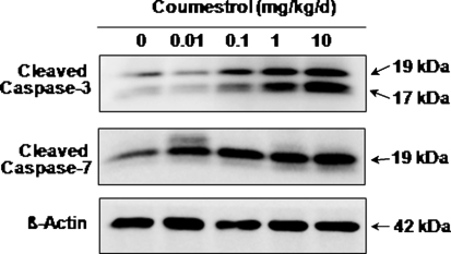
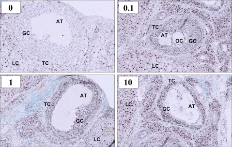
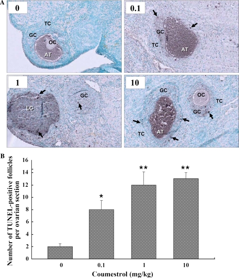
This study is the first to examine the increased apoptosis in the adult rat ovary after lactational exposure to coumestrol (COU), a potent phytoestrogen. Lactating dams were gavaged at doses of 0.01, 0.1, 1, and 10 mg/kg COU during the lactation period and the reproductive effects of female pups were investigated in young adults. Rats were sacrificed at postnatal days (PND) 81-84. Ovarian weights were reduced significantly at 0.1 and 1.0 mg/kg COU. The reduction in the ovarian weight occurred in parallel with an increase in the apoptosis at PND 135-140. A marked dose-dependent increase in the expressions of active caspase-3 and -7 was observed in ovarian granulosa cells. Immunostaining for active caspase-3 and the TUNEL staining of apoptotic cells were also increased in ovaries exposed to COU in a dose-dependent manner. These results suggest new sights into the effect of lactational exposure to COU on the female reproductive health.
Figures
References
-
- None
- Adlercreutz H, Mazur W (1998) Overview of naturally occurring endocrine-active substances in the human diet. In: Dunaif GE, Olin SS, Scimeca JA, Thomas JA (eds) Human diet and endocrine modulation. ILSI Press, Washington, DC, pp 134–285
-
- {'text': '', 'ref_index': 1, 'ids': [{'type': 'PubMed', 'value': '1659780', 'is_inner': True, 'url': 'https://pubmed.ncbi.nlm.nih.gov/1659780/'}]}
- Adlercreutz H, Honjo H, Higashi A, Fotsis T, Hämäläinen E, Hasegawa T, Okada H (1991) Urinary excretion of lignans and isoflavonoids phytoestrogens in Japanese men and women consuming a traditional Japanese diet. Am J Clin Nutr 54:1093–1100 - PubMed
-
- {'text': '', 'ref_index': 1, 'ids': [{'type': 'PubMed', 'value': '2019275', 'is_inner': True, 'url': 'https://pubmed.ncbi.nlm.nih.gov/2019275/'}]}
- Bendell JJ, Dorrington J (1991) 17β-Estradiol stimulates DNA synthesis in rat granulosa cells: action mediated by transforming growth factor-p. Endocrinology 128:2663–2665 - PubMed
-
- {'text': '', 'ref_index': 1, 'ids': [{'type': 'DOI', 'value': '10.1210/en.133.5.2204', 'is_inner': False, 'url': 'https://doi.org/10.1210/en.133.5.2204'}, {'type': 'PubMed', 'value': '8404672', 'is_inner': True, 'url': 'https://pubmed.ncbi.nlm.nih.gov/8404672/'}]}
- Billig H, Furuta I, Hsueh AJ (1993) Estrogens inhibit and androgens enhance ovarian granulosa cell apoptosis. Endocrinology 133:2204–2212 - PubMed
-
- {'text': '', 'ref_index': 1, 'ids': [{'type': 'DOI', 'value': '10.1095/biolreprod57.4.813', 'is_inner': False, 'url': 'https://doi.org/10.1095/biolreprod57.4.813'}, {'type': 'PubMed', 'value': '9314585', 'is_inner': True, 'url': 'https://pubmed.ncbi.nlm.nih.gov/9314585/'}]}
- Boone DL, Tsang BK (1997) Identification and localization of DNase I in the rat ovary. Biol Reprod 57:813–821 - PubMed
Publication types
MeSH terms
Substances
LinkOut - more resources
Full Text Sources
Research Materials
