. 2009 May;14(4):507-19.
doi: 10.1007/s00775-009-0466-z. Epub 2009 Jan 23.

Auranofin disrupts selenium metabolism in Clostridium difficile by forming a stable Au-Se adduct

Affiliations

Auranofin disrupts selenium metabolism in Clostridium difficile by forming a stable Au-Se adduct

Sarah Jackson-Rosario et al. J Biol Inorg Chem. 2009 May.

Abstract

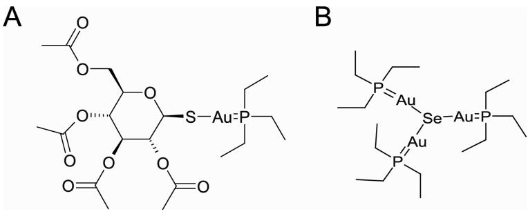
Clostridium difficile is a nosocomial pathogen whose incidence and importance are on the rise. Previous work in our laboratory characterized the central role of selenoenzyme-dependent Stickland reactions in C. difficile metabolism. In this work we have identified, using mass spectrometry, a stable complex formed upon reaction of auranofin (a gold-containing drug) with selenide in vitro. X-ray absorption spectroscopy supports the structure that we proposed on the basis of mass-spectrometric data. Auranofin potently inhibits the growth of C. difficile but does not similarly affect other clostridia that do not utilize selenoproteins to obtain energy. Moreover, auranofin inhibits the incorporation of radioisotope selenium ((75)Se) in selenoproteins in both Escherichia coli, the prokaryotic model for selenoprotein synthesis, and C. difficile without impacting total protein synthesis. Auranofin blocks the uptake of selenium and results in the accumulation of the auranofin-selenide adduct in the culture medium. Addition of selenium in the form of selenite or L-selenocysteine to the growth medium significantly reduces the inhibitory action of auranofin on the growth of C. difficile. On the basis of these results, we propose that formation of this complex and the subsequent deficiency in available selenium for selenoprotein synthesis is the mechanism by which auranofin inhibits C. difficile growth. This study demonstrates that targeting selenium metabolism provides a new avenue for antimicrobial development against C. difficile and other selenium-dependent pathogens.


Figures

Figure 1
Figure 1. HPLC analysis of products formed during reaction of auranofin and hydrogen selenide
Auranofin (10 mM in DMSO) was mixed anaerobically with equal volumes of H2O (a) or 20 mM hydrogen selenide (b). Reactions were incubated for 20 minutes before injection. 20 μL samples were loaded onto a C18 column at a flow rate of 0.5 mL per minute. The starting solvent (used for injection) was 0.05% trifluoroacetic (TFA) acid in H2O. A linear gradient (50 minutes) was developed to 100% acetonitrile, 0.05% TFA. Products were identified at 254 nm using a diode-array detector.
Figure 2
Figure 2. Mass spectrometry reveals major reaction products of auranofin and hydrogen selenide
The predominant product from direct injection of an equimolar reaction of auranofin and hydrogen selenide has a mass of 1025.10 atomic mass units and exhibits a selenium isotope signature [51]. The inset plot shows all of the products obtained across the entire spectrum. The predominant compound was also obtained after liquid chromatography as described in the methods section, and its mass was confirmed (data not shown).
Figure 3
Figure 3
Structure of auranofin (A) and the putative structure of the stable product formed upon reaction of auranofin and selenide (B), based on mass spectrometry
Figure 4
Figure 4. Au L3 XAS Spectra
Au L3 edge XAS spectra for Au(III) compounds (KAuBr4, dashed black; KAuCl4, dashed red), Au(I) compound (Na3Au(S2O3)2, dashed green), compared to that of auranofin (solid black) and the product of reaction of auranofin with hydrogen selenide (solid red).
Figure 5
Figure 5. Curve-fitting analysis of Au L3 (top) and Se K (bottom) EXAFS data for the product of the reaction of auranofin with hydrogen selenide
In all figures, thin black lines represent the observed data (Fourier transforms, FT, in full frames, EXAFS in insets) and bold red lines represent the best-fit simulation using feff v7 calculations based on molecular models derived from the hypothesized structure in Figure 3B. The model used for Au EXAFS consisted of a P–Au–Se fragment with a P-Au-Se angle of 180°; the simulation was generated by the parameters summarized in Fit 5 of Table 1. The model use for Se EXAFS consisted of a Se surrounded by three Au–P fragments with Au directly bonded to Se, Se-Au-P angles of 180° and Au-Se-Au angles of 120°; the simulation was generated by the parameters summarized in Fit 6 of Table R. Fourier transforms were k3 weighted over the k = 2.0 – 13.0 Å−1 range (Au L3) or the k = 2.0 – 14.0 Å−1 (Se K) range and were phase-corrected based on P.
Figure 6
Figure 6. Auranofin inhibits growth of C. difficile
C. difficile cultures (NAP1/O27, VPI10463, 630, and ATCC 9689) were grown anaerobically in BHI + cysteine. Auranofin in DMSO (0.25, 0.5, 0.75, 0.8, 0.85, 0.9, 0.95, 1.0, 1.5, and 2.0 μM) was added to the growth medium prior to inoculation. Growth was measured as optical density at 600 nm after 24 hours at 37°C. Percent growth (growth yield of inhibited cultures versus control) is plotted at the indicated concentrations of auranofin. Data shown is from at least three independent cultures. Error bars indicate standard deviation.
Figure 7
Figure 7. Auranofin prevents incorporation of 75Se in anaerobically grown E. coli and C. difficile
Soluble extracts of MC4100 treated with varying concentrations of auranofin (0, 5, 10, 50 μM lanes 1-4 respectively) were prepared following anaerobic growth overnight in the presence of 6 μCi of 75Se (0.1 μM Na2SeO3) (a) or 20 μCi of 35S (b). Twenty-five micrograms of protein from crude cell extracts were separated by a 12% SDS-polyacrylamide gel, and the radioactive bands were visualized using a phosphoimager. (c). Soluble extracts of C. difficile (NAPI/O27 strain) were prepared after treatment mid-logarithmic phase with auranofin (0, 5, 10 μM lanes 1-3 respectively) in the presence of 6 μCi of 75Se (0.1 μM Na2SeO3) (a) or 20 μCi of 35S (d). Twenty-five micrograms of protein from crude cell extracts were separated by a 12% SDS-polyacrylamide gel, and the radioactive bands were visualized using a phosphoimager. The predicted selenoproteins are indicated by arrows based on the molecular weights of the genes as described previously [17].
Figure 8
Figure 8. Auranofin prevents uptake of 75Se by E. coli and C. difficile
Actively growing cultures of E. coli (A) and C. difficile(B) were treated with auranofin (0 to 25 μM as indicated) as well as 75Se. Cultures were incubated for 20 minutes at 37°C. Cells were harvested by centrifugation and washed with PBS. Total uptake of 75Se was analyzed using a Model 1470 Gamma counter (Perkin–Elmer, Wellesley, ME) and is reported as counts per minute (CPM). Data shown is from at least three independent cultures. Error bars indicate standard dEviation.
Figure 9
Figure 9. Auranofin forms a complex with radiolabeled selenium in C. difficile growth medium
Growth media from the uptake experiments (0 and 10μM auranofin, a and b respectively) were separated by HPLC as described for the analysis of the mixtures of auranofin and hydrogen selenide. Fractions were collected every minute and analyzed using the gamma counter
Figure 10
Figure 10. Selenium supplementation prevents auranofin-dependent growth inhibition of C. difficile
C. difficile (NAPI/O27) was cultivated in BHI + cysteine. The culture medium was supplemented with either sodium selenite (0.5, 1.0, 5.0 μM) (a) or L-selenocysteine (0.5, 1.0, 5.0 μM) (b). Percent growth (growth yield of inhibited cultures versus control) is plotted at the indicated concentrations of auranofin after 24 hours at 37°C. Data shown is from at least three independent cultures. Error bars indicate standard deviation.

References

    1. Kean WF, Hart L, Buchanan WW. British journal of rheumatology. 1997;36:560–572. - PubMed
    1. Messori L, Marcon G. Met Ions Biol Syst. 2004;41:279–304. - PubMed
    1. Becker K, Gromer S, Schirmer RH, Muller S. Eur J Biochem. 2000;267:6118–6125. - PubMed
    1. Gromer S, Arscott LD, Williams CH, Jr., Schirmer RH, Becker K. J Biol Chem. 1998;273:20096–20101. - PubMed
    1. Lobanov AV, Gromer S, Salinas G, Gladyshev VN. Nucleic Acids Res. 2006;34:4012–4024. - PMC - PubMed

Publication types

MeSH terms

LinkOut - more resources