

Random dissection to select for protein split sites and its application in protein fragment complementation
- PMID: 19165722
- PMCID: PMC2708047
- DOI: 10.1002/pro.42
Random dissection to select for protein split sites and its application in protein fragment complementation
Abstract
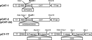
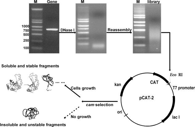
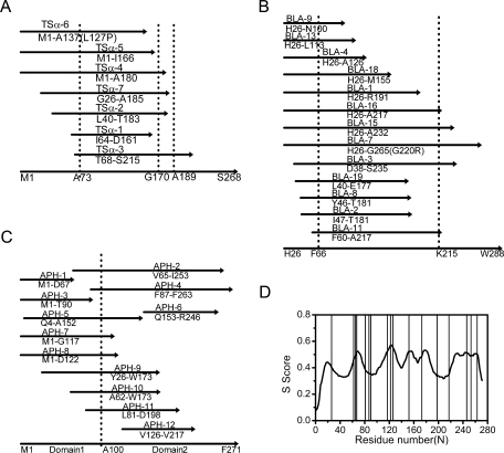
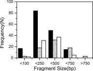
To identify protein split sites quickly, a selection procedure by using chloramphenicol acetyl transferase (CAT) as reporter was introduced to search for folded protein fragments from libraries generated by random digestion and reassembly of the target gene, which yielded an abundant amount of DNA fragments with controllable lengths. Experimental results of tryptophan synthase alpha subunit (TSalpha) and TEM-1 beta-lactamase agreed well with what the literature has reported. The solubility of these fragments correlated roughly with the minimum inhibitory concentrations of the CAT fusions. The application of this dissection protocol to protein fragment complementation assay (PCA) was evaluated using aminoglycoside-3'-phosphotransferase I (APH(3')-I) as a model protein. Three nearly bisectional sites and a number of possible split points were identified, and guided by this result, four novel pairs of fragments were tested for complementation. Three out of four pairs partially restored the APH activity with the help of leucine zippers, and a truncated but active APH(3')-I (Delta1-25) was also found. Finally, the weakly active APH(3')-I-(1-253)NZ/CZ (254-271) containing a short 18 residue tag was further improved by error-prone PCR, and a best mutant was obtained showing a fourfold improvement after just one round of evolution. These results demonstrate that protein random dissection based on the CAT selection can provide an efficient search for protein breakage points and guide the design of fragments for protein complementation assay. Furthermore, more active fragment pairs can be achieved with the classical directed evolution approach.
Figures
Similar articles
-
Enhanced catalytic efficiency of aminoglycoside phosphotransferase (3')-IIa achieved through protein fragmentation and reassembly.J Mol Biol. 2005 Oct 14;353(1):26-37. doi: 10.1016/j.jmb.2005.08.026. J Mol Biol. 2005. PMID: 16168439
-
Mapping of unit boundaries of a protein: exhaustive search for permissive sites for duplication by complementation analysis of random fragment libraries of tryptophan synthase alpha subunit.J Mol Biol. 2004 Jan 23;335(4):1093-104. doi: 10.1016/j.jmb.2003.11.029. J Mol Biol. 2004. PMID: 14698302
-
Beta-lactamase protein fragment complementation assays as in vivo and in vitro sensors of protein protein interactions.Nat Biotechnol. 2002 Jun;20(6):619-22. doi: 10.1038/nbt0602-619. Nat Biotechnol. 2002. PMID: 12042868
-
Antibody library selection by the {beta}-lactamase protein fragment complementation assay.Protein Eng Des Sel. 2009 Mar;22(3):149-58. doi: 10.1093/protein/gzn053. Epub 2008 Sep 30. Protein Eng Des Sel. 2009. PMID: 18829449
-
Selection of TNF-alpha binding affibody molecules using a beta-lactamase protein fragment complementation assay.N Biotechnol. 2009 Nov 30;26(5):251-9. doi: 10.1016/j.nbt.2009.06.980. Epub 2009 Jul 1. N Biotechnol. 2009. PMID: 19576305
Cited by
-
A 'resource allocator' for transcription based on a highly fragmented T7 RNA polymerase.Mol Syst Biol. 2014 Jul 30;10(7):742. doi: 10.15252/msb.20145299. Mol Syst Biol. 2014. PMID: 25080493 Free PMC article.
-
Design of split superantigen fusion proteins for cancer immunotherapy.J Biol Chem. 2019 Apr 19;294(16):6294-6305. doi: 10.1074/jbc.RA118.006742. Epub 2019 Feb 19. J Biol Chem. 2019. PMID: 30782846 Free PMC article.
-
Complementation between inactive fragments of SssI DNA methyltransferase.BMC Mol Biol. 2012 May 30;13:17. doi: 10.1186/1471-2199-13-17. BMC Mol Biol. 2012. PMID: 22646482 Free PMC article.
References
-
- Ladurner A, Itzhaki L, Gay G, Fersht A. Complementation of peptide fragments of the single domain protein chymotrypsin inhibitor 2. J Mol Biol. 1997;273:317–329. - PubMed
-
- Hayes F, Hallet B, Cao YH. Insertion mutagenesis as a tool in the modification of protein function. J Biol Chem. 1997;272:28833–28836. - PubMed
-
- Voigt CA, Martinez C, Wang ZG, Mayo SL, Arnold FH. Protein building blocks preserved by recombination. Nat Struct Biol. 2002;9:553–558. - PubMed
Publication types
MeSH terms
Substances
LinkOut - more resources
Full Text Sources
Other Literature Sources
Miscellaneous
