. 2009 Feb;18(2):424-33.
doi: 10.1002/pro.45.

Methionine oxidation in human IgG2 Fc decreases binding affinities to protein A and FcRn

Affiliations

Methionine oxidation in human IgG2 Fc decreases binding affinities to protein A and FcRn

Hai Pan et al. Protein Sci. 2009 Feb.

Abstract

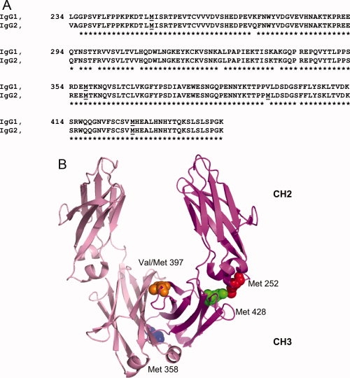
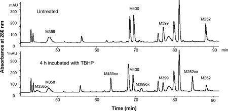
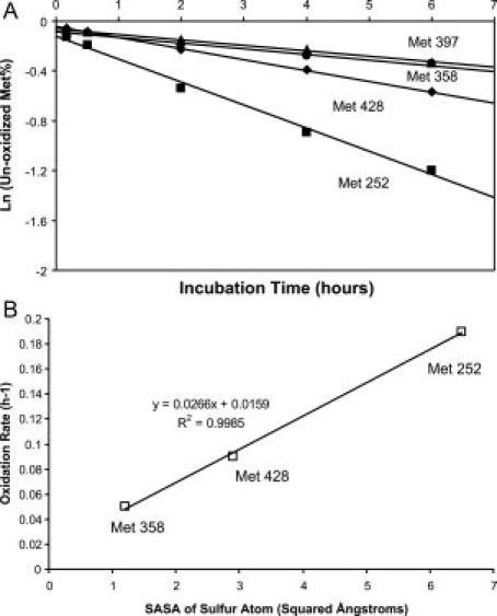
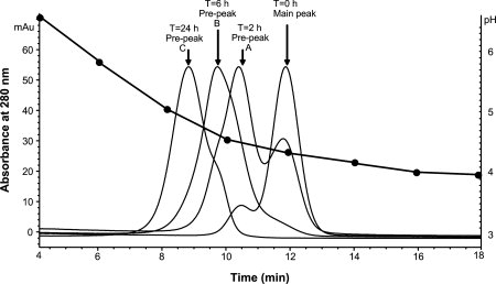
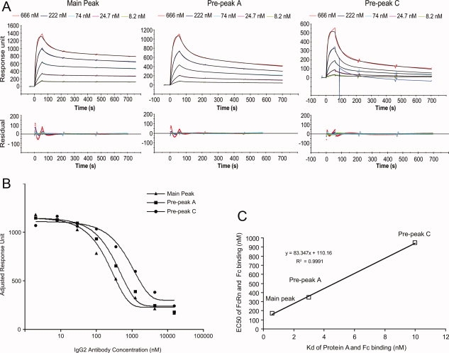
Susceptibility of methionine residues to oxidation is a significant issue of protein therapeutics. Methionine oxidation may limit the product's clinical efficacy or stability. We have studied kinetics of methionine oxidation in the Fc portion of the human IgG2 and its impact on the interaction with FcRn and Protein A. Our results confirm previously published observations for IgG1 that two analogous solvent-exposed methionine residues in IgG2, Met 252 and Met 428, oxidize more readily than the other methionine residue, Met 358, which is buried inside the Fc. Met 397, which is not present in IgG1 but in IgG2, oxidizes at similar rate as Met 358. Oxidation of two labile methionines, Met 252 and Met 428, weakens the binding of the intact antibody with Protein A and FcRn, two natural protein binding partners. Both of these binding partners share the same binding site on the Fc. Additionally, our results shows that Protein A may serve as a convenient and inexpensive surrogate for FcRn binding measurements.


Figures

Figure 1
Figure 1
A: Alignment of the amino acid sequences of human IgG1 and IgG2 Fc. Residues are numbered using the EU numbering system. Methionine residues are underlined. B: The cartoon representation of the Fc. The crystal structure of human IgG1 Fc (pdb code:1FC1) was used to prepare the figure. Met 252 was colored red. Met 358 was colored blue. Val 397, which is equivalent to Met 397 in human IgG2, was colored orange and Met 428 was colored green. The sphere presentations of these residues are only shown in one peptide chain of the Fc. [Color figure can be viewed in the online issue, which is available at www.interscience.wiley.com.]:
Figure 2
Figure 2
Peptide maps for nonreduced Lys-C digests for the untreated IgG2 and treated IgG2 that has been incubated with TBHP for 4 h.
Figure 3
Figure 3
A: Kinetics of methionine oxidation. Oxidation rates of each methionine (square for Met 252, diamond for Met 428, circle for Met 358, and triangle for Met 397) were determined from the slopes of the linear fits through the data. B: The correlation between oxidation rates and solvent accessible surface area of sulfur atom for each methionine.
Figure 4
Figure 4
pH-gradient Protein A chromatograms of an IgG2 antibody treated by TBHP for various time periods. The pH gradient was superimposed. Each filled circle represents the pH value of the eluent collected in every 2 min.
Figure 5
Figure 5
A: The sensorgrams (colored lines) of the main peak, prepeak A, and prepeak C binding with Protein A immobilized on the Biacore chip. In each panel, the fitted curves are colored black. A spike was observed in the sensorgram of injecting 222 nM prepeak C to the Biacore chip probably due to an air bubble. The curve of this sensorgram was fitted before the spike and the residuals weren't shown after the spike. B: The competition binding curves of the main peak, prepeak A, and prepeak C with FcRn. C: The correlation between Kd of Fc and Protein A and the EC50 value of Fc and FcRn for main peak, prepeak A, and prepeak C. [Color figure can be viewed in the online issue, which is available at www.interscience.wiley.com.]:

References

    1. Shacter E. Quantification and significance of protein oxidation in biological samples. Drug Metab Rev. 2000;32:307–326. - PubMed
    1. Scislowski PW, Foster AR, Fuller MF. Regulation of oxidative degradation of l-lysine in rat liver mitochondria. Biochem J. 1994;300(Pt 3):887–891. - PMC - PubMed
    1. Hovorka S, Schoneich C. Oxidative degradation of pharmaceuticals: theory, mechanisms and inhibition. J Pharm Sci. 2001;90:253–269. - PubMed
    1. Slavica A, Dib I, Nidetzky B. Single-site oxidation, cysteine 108 to cysteine sulfinic acid, in D-amino acid oxidase from Trigonopsis variabilis and its structural and functional consequences. Appl Environ Microbiol. 2005;71:8061–8068. - PMC - PubMed
    1. Lu HS, Fausset PR, Narhi LO, Horan T, Shinagawa K, Shimamoto G, Boone TC. Chemical modification and site-directed mutagenesis of methionine residues in recombinant human granulocyte colony-stimulating factor: effect on stability and biological activity. Arch Biochem Biophys. 1999;362:1–11. - PubMed

MeSH terms

LinkOut - more resources