Unique interactions between the chromophore and glutamate 16 lead to far-red emission in a red fluorescent protein
- PMID: 19165727
- PMCID: PMC2708057
- DOI: 10.1002/pro.66
Unique interactions between the chromophore and glutamate 16 lead to far-red emission in a red fluorescent protein
Abstract
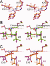
mPlum is a far-red fluorescent protein with emission maximum at approximately 650 nm and was derived by directed evolution from DsRed. Two residues near the chromophore, Glu16 and Ile65, were previously revealed to be indispensable for the far-red emission. Ultrafast time-resolved fluorescence emission studies revealed a time dependent shift in the emission maximum, initially about 625 nm, to about 650 nm over a period of 500 ps. This observation was attributed to rapid reorganization of the residues solvating the chromophore within mPlum. Here, the crystal structure of mPlum is described and compared with those of two blue shifted mutants mPlum-E16Q and -I65L. The results suggest that both the identity and precise orientation of residue 16, which forms a unique hydrogen bond with the chromophore, are required for far-red emission. Both the far-red emission and the time dependent shift in emission maximum are proposed to result from the interaction between the chromophore and Glu16. Our findings suggest that significant red shifts might be achieved in other fluorescent proteins using the strategy that led to the discovery of mPlum.
Figures


Similar articles
-
Extended Stokes shift in fluorescent proteins: chromophore-protein interactions in a near-infrared TagRFP675 variant.Sci Rep. 2013;3:1847. doi: 10.1038/srep01847. Sci Rep. 2013. PMID: 23677204 Free PMC article.
-
On the Nature of an Extended Stokes Shift in the mPlum Fluorescent Protein.J Phys Chem B. 2015 Oct 15;119(41):13052-62. doi: 10.1021/acs.jpcb.5b07724. Epub 2015 Oct 2. J Phys Chem B. 2015. PMID: 26402581
-
Recovery of red fluorescent protein chromophore maturation deficiency through rational design.PLoS One. 2012;7(12):e52463. doi: 10.1371/journal.pone.0052463. Epub 2012 Dec 20. PLoS One. 2012. PMID: 23285050 Free PMC article.
-
Red fluorescent proteins: chromophore formation and cellular applications.Curr Opin Struct Biol. 2012 Oct;22(5):679-88. doi: 10.1016/j.sbi.2012.09.002. Epub 2012 Sep 20. Curr Opin Struct Biol. 2012. PMID: 23000031 Free PMC article. Review.
-
Chromophore transformations in red fluorescent proteins.Chem Rev. 2012 Jul 11;112(7):4308-27. doi: 10.1021/cr2001965. Epub 2012 May 4. Chem Rev. 2012. PMID: 22559232 Free PMC article. Review. No abstract available.
Cited by
-
Generation of longer emission wavelength red fluorescent proteins using computationally designed libraries.Proc Natl Acad Sci U S A. 2010 Nov 23;107(47):20257-62. doi: 10.1073/pnas.1013910107. Epub 2010 Nov 8. Proc Natl Acad Sci U S A. 2010. PMID: 21059931 Free PMC article.
-
Red fluorescent proteins: advanced imaging applications and future design.Angew Chem Int Ed Engl. 2012 Oct 22;51(43):10724-38. doi: 10.1002/anie.201200408. Epub 2012 Jul 31. Angew Chem Int Ed Engl. 2012. PMID: 22851529 Free PMC article. Review.
-
Genetically encoding new bioreactivity.N Biotechnol. 2017 Sep 25;38(Pt A):16-25. doi: 10.1016/j.nbt.2016.10.003. Epub 2016 Oct 6. N Biotechnol. 2017. PMID: 27721014 Free PMC article. Review.
-
Distinct effects of guanidine thiocyanate on the structure of superfolder GFP.PLoS One. 2012;7(11):e48809. doi: 10.1371/journal.pone.0048809. Epub 2012 Nov 7. PLoS One. 2012. PMID: 23144981 Free PMC article.
-
Extended Stokes shift in fluorescent proteins: chromophore-protein interactions in a near-infrared TagRFP675 variant.Sci Rep. 2013;3:1847. doi: 10.1038/srep01847. Sci Rep. 2013. PMID: 23677204 Free PMC article.
References
-
- Weissleder R, Ntziachristos V. Shedding light onto live molecular targets. Nat Med. 2003;9:123–128. - PubMed
-
- Gurskaya N, Fradkov A, Terskikh A, Matz M, Labas Y, Martynov V, Yanushevich Y, Lukyanov K, Lukyanov S. GFP-like chromoproteins as a source of far-red fluorescent proteins. FEBS lett. 2001;507:16–20. - PubMed
Publication types
MeSH terms
Substances
LinkOut - more resources
Full Text Sources

