. 2010 Jan;92(1):18-32.
doi: 10.1002/jbm.a.32279.

A bovine acellular scaffold for vocal fold reconstruction in a rat model

Affiliations

A bovine acellular scaffold for vocal fold reconstruction in a rat model

Chet C Xu et al. J Biomed Mater Res A. 2010 Jan.

Abstract

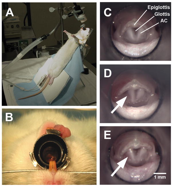
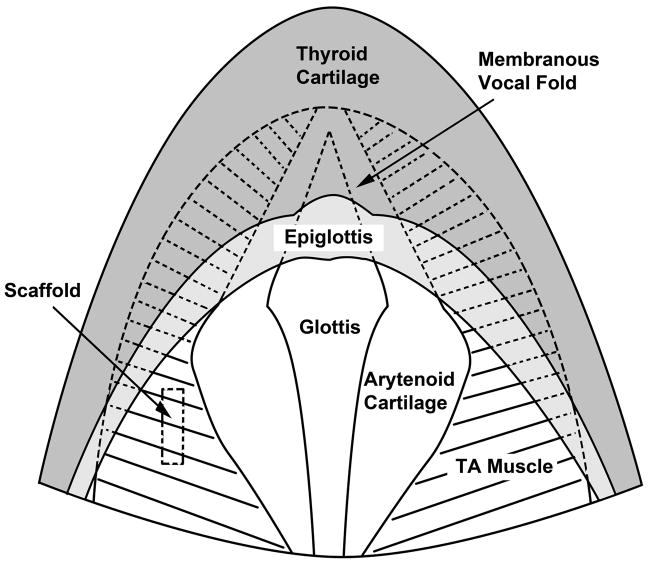
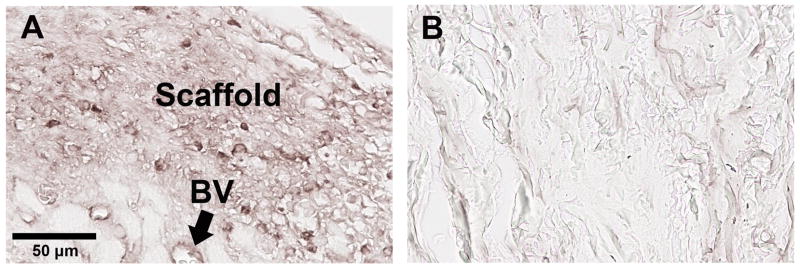
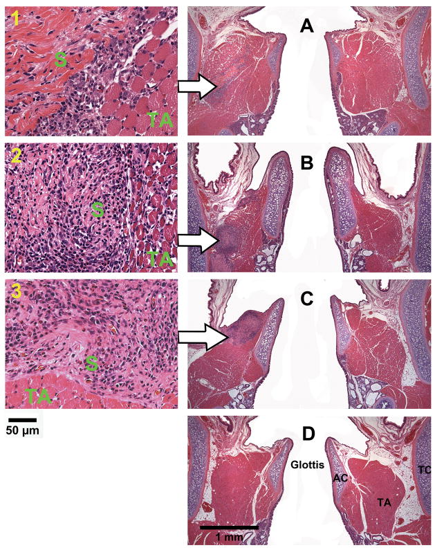
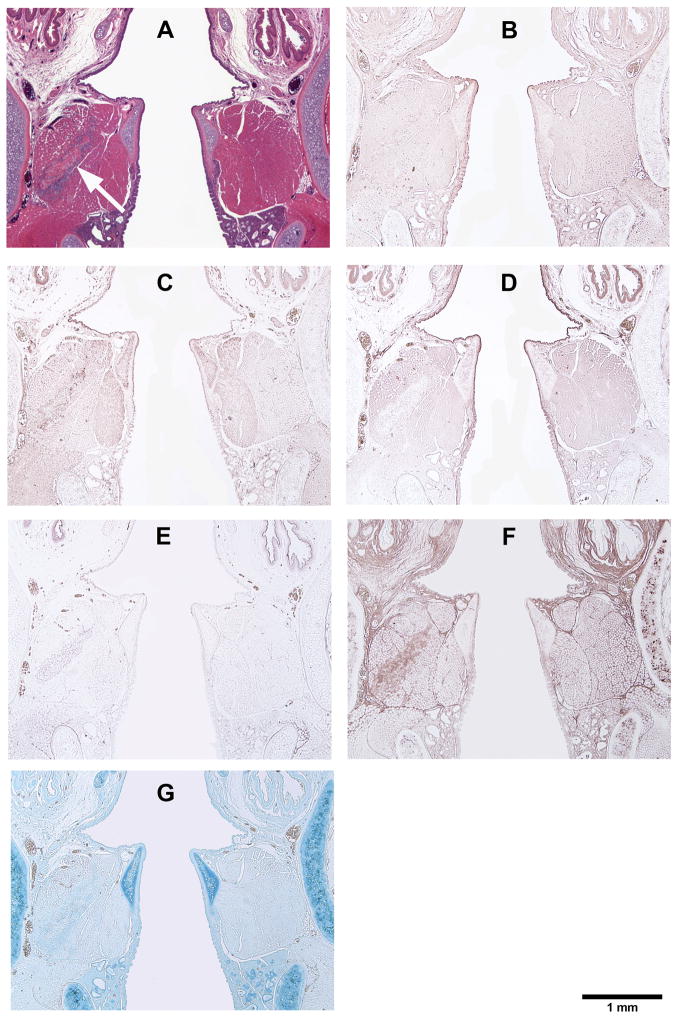
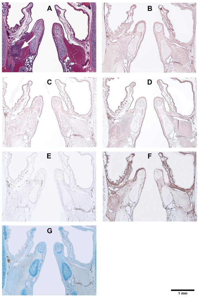
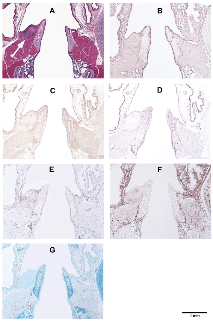
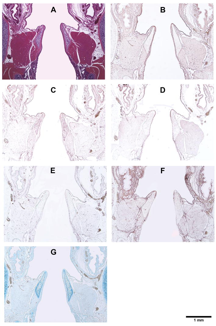
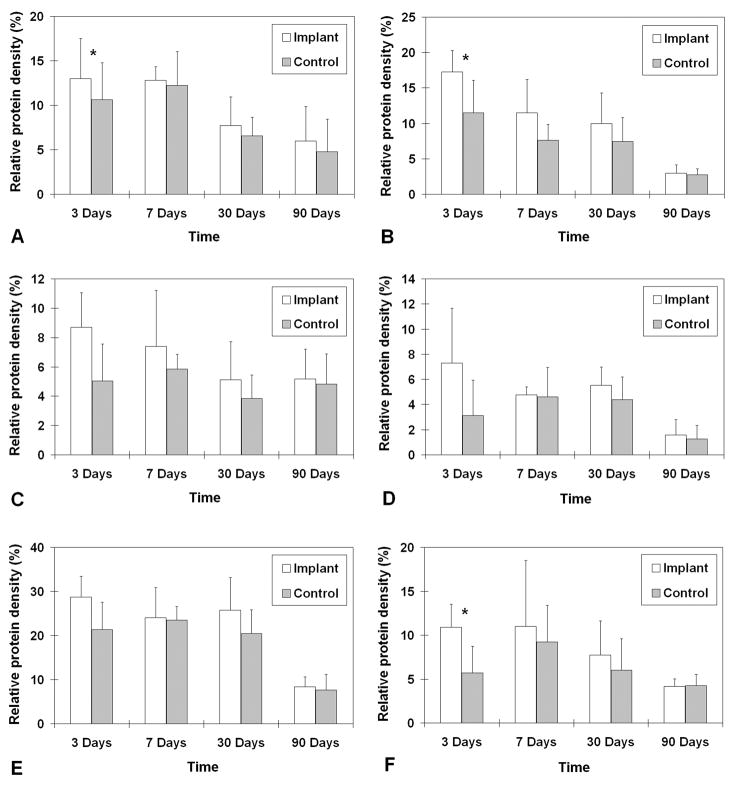
With a rat model of vocal fold injury, this study examined the in vivo host response to an acellular xenogeneic scaffold derived from the bovine vocal fold lamina propria, and the potential of the scaffold for constructive tissue remodeling. Bilateral wounds were created in the posterior vocal folds of 20 rats, and bovine acellular scaffolds were implanted into the wounds unilaterally, with the contralateral vocal folds as control. The rats were humanely sacrificed after 3 days, 7 days, 1 month, and 3 months, and the coronal sections of their larynges were examined histologically. Expressions of key matrix proteins including collagen I, collagen III, elastin, fibronectin, hyaluronic acid, and glycosaminoglycans (GAGs) were quantified with digital image analysis. Significant infiltration of host inflammatory cells and host fibroblasts in the scaffold implant was observed in the acute stage of wound repair (3 days and 7 days postsurgery). The mean relative densities of collagen I, collagen III, and GAGs in the implanted vocal folds were significantly higher than those in the control after 3 days, followed by gradual decreases over 3 months. Histological results showed that the scaffolds were apparently degraded by 3 months, with no fibrotic tissue formation or calcification. These preliminary findings suggested that the bovine acellular scaffold could be a potential xenograft for vocal fold regeneration.


Figures

Figure 1
Figure 1
Illustration of the surgical procedure for vocal fold implantation in the rat model. (A) A rat was secured on a customized surgical platform in supine position. (B) The rat larynx was visualized with a surgical microscope through a 4.0-mm otoscope specula. (C) Superior view of the rat larynx through the surgical microscope (top: anterior; bottom: posterior). (D) A wound was surgically created by a 1-mm long longitudinal incision on the superior surface of the left, posterior vocal fold (arrow) and two bovine acellular scaffold samples were implanted into the wound. No wound was created on the contralateral (right) vocal fold for this particular rat, but wounds were created on the right vocal fold as control in 20 rats. (E) 24 hours following surgery, the vocal fold implanted with the scaffolds (arrow) appeared normal (AC = arytenoid cartilage)
Figure 2
Figure 2
Schematic of the anatomy of the rat larynx as seen from a superior view similar to that of Figure 1C. The membranous vocal folds (shown in dotted lines) are obstructed by the epiglottis at this angle of view. The approximate location of an implanted scaffold is shown in the left cartilaginous vocal fold (TA = thyroarytenoid muscle).
Figure 3
Figure 3
Immuno-histochemical examination of fibronectin in (A) rat laryngeal coronal section; (B) bovine acellular scaffold section. The rat laryngeal specimen was harvested 30 days after the bovine scaffold implantation, showing the posterior vocal fold mucosa. The anti-rat fibronectin antibody reacts positively with the laryngeal mucosa integrated with the implanted scaffold in (A) (Left: medial; Right: lateral), whereas only background stain is observed for the bovine acellular scaffold in (B) (BV = blood vessels).
Figure 4
Figure 4
Histological coronal sections of rat laryngeal specimens stained with H&E, showing the acellular scaffolds implanted into the left vocal fold wounds (A) 3 days, (B) 7 days, (C) 30 days, and (D) 90 days after surgery (total magnification = 40×). Arrows indicate the implants in the left vocal folds (experimental vocal folds). Images on the left are at higher magnification (400×) showing the interface between the scaffold and the host tissue (1) 3 days, (2) 7 days, and (3) 30 days after surgery (AC = arytenoid cartilage; TA = thyroarytenoid muscle; TC = thyroid cartilage; S = implanted acellular scaffold).
Figure 5
Figure 5
Histological coronal sections of rat laryngeal specimens showing the acellular scaffolds implanted into the left vocal fold wounds 3 days after surgery, with the right vocal fold wounds as control: (A) H&E (arrow indicates the implant in the left vocal fold); (B) Collagen type I; (C) Collagen type III; (D) Elastin; (E) Fibronectin; (F) Hyaluronic acid; (G) Glycosaminoglycans.
Figure 6
Figure 6
Histological coronal sections of rat laryngeal specimens showing the acellular scaffolds implanted into the left vocal fold wounds 7 days after surgery, with the right vocal fold wounds as control: (A) H&E (arrow indicates the implant in the left vocal fold); (B) Collagen type I; (C) Collagen type III; (D) Elastin; (E) Fibronectin; (F) Hyaluronic acid; (G) Glycosaminoglycans.
Figure 7
Figure 7
Histological coronal sections of rat laryngeal specimens showing the acellular scaffolds implanted into the left vocal fold wounds 30 days after surgery, with the right vocal fold wounds as control: (A) H&E (arrow indicates the implant in the left vocal fold); (B) Collagen type I; (C) Collagen type III; (D) Elastin; (E) Fibronectin; (F) Hyaluronic acid; (G) Glycosaminoglycans.
Figure 8
Figure 8
Histological coronal sections of rat laryngeal specimens showing the acellular scaffolds implanted into the left vocal fold wounds 90 days after surgery, with the right vocal fold wounds as control: (A) H&E (note the absence of the implant in the left vocal fold); (B) Collagen type I; (C) Collagen type III; (D) Elastin; (E) Fibronectin; (F) Hyaluronic acid; (G) Glycosaminoglycans.
Figure 9
Figure 9
Relative densities of ECM proteins in the rat vocal fold implanted with the bovine acellular scaffold versus those in the control vocal fold (n = 5): (A) Collagen type I; (B) Collagen type III; (C) Elastin; (D) Fibronectin; (E) Hyaluronic acid; (F) Glycosaminoglycans. * p < 0.05 for differences between the implanted vocal fold and the control.

Similar articles

Cited by

References

    1. Ramig LO, Verdolini K. Treatment efficacy: voice disorders. J Speech Lang Hear Res. 1998;41:S101–116. - PubMed
    1. Hirano S. Current treatment of vocal fold scarring. Curr Opin Otolaryngol Head Neck Surg. 2005;13:143–147. - PubMed
    1. Gray SD, Titze IR, Chan RW, Hammond TH. Vocal fold proteoglycans and their influence on biomechanics. Laryngoscope. 1999;109:845–854. - PubMed
    1. Gray SD, Titze IR, Alipour F, Hammond TH. Biomechanical and histologic observations of vocal fold fibrous proteins. Ann Otol Rhinol Laryngol. 2000;109:77–85. - PubMed
    1. Sato K, Hirano M, Nakashima T. Stellate cells in the human vocal fold. Ann Otol Rhinol Laryngol. 2001;110:319–325. - PubMed

Publication types

LinkOut - more resources