Review
. 2009 Jan;214(1):1-18.
doi: 10.1111/j.1469-7580.2008.01011.x.

The fascia of the limbs and back--a review

Affiliations
Review

The fascia of the limbs and back--a review

Mike Benjamin. J Anat. 2009 Jan.

Abstract

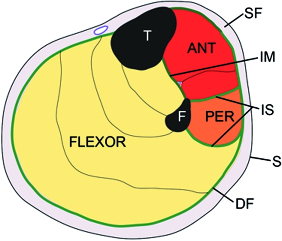
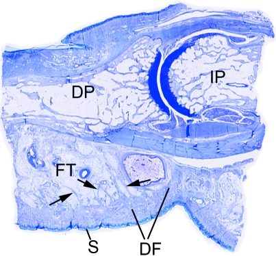
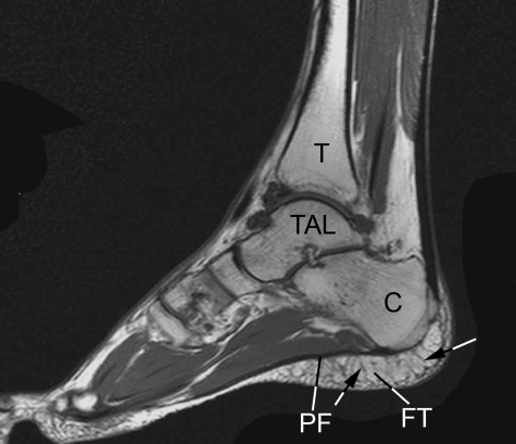
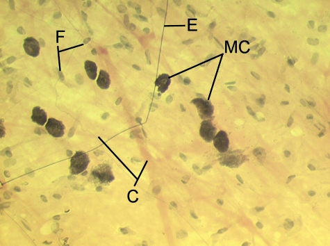
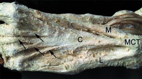
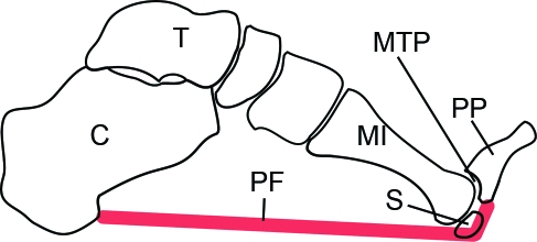
Although fasciae have long interested clinicians in a multitude of different clinical and paramedical disciplines, there have been few attempts to unite the ensuing diverse literature into a single review. The current article gives an anatomical perspective that extends from the gross to the molecular level. For expediency, it deals only with fascia in the limbs and back. Particular focus is directed towards deep fascia and thus consideration is given to structures such as the fascia lata, thoracolumbar fascia, plantar and palmar fascia, along with regional specializations of deep fascia such as retinacula and fibrous pulleys. However, equal emphasis is placed on general aspects of fascial structure and function, including its innervation and cellular composition. Among the many functions of fascia considered in detail are its ectoskeletal role (as a soft tissue skeleton for muscle attachments), its importance for creating osteofascial compartments for muscles, encouraging venous return in the lower limb, dissipating stress concentration at entheses and acting as a protective sheet for underlying structures. Emphasis is placed on recognizing the continuity of fascia between regions and appreciating its key role in coordinating muscular activity and acting as a body-wide proprioceptive organ. Such considerations far outweigh the significance of viewing fascia in a regional context alone.


Figures

Fig. 1
Fig. 1
A diagrammatic representation of a transverse section through the upper part of the leg showing the relative positions of the superficial (SF) and deep fascia (DF) in relation to the skin (S) and muscles. Note how the deep fascia, in association with the bones [tibia (T) and fibula (F)] and intermuscular septa (IS) forms a series of osteofascial compartments housing the extensor, peroneal (PER) and flexor muscles. If pressure builds up within a compartment because of an acute or overuse injury, then the vascular supply to the muscles within it can be compromised and ischaemia results. ANT, anterior compartment; IM, interosseous membrane.
Fig. 2
Fig. 2
A low power view of a sagittal section through a finger in the region of the distal interphalangeal joint. Note how the skin (S) on the palmar side is intimately associated with a thick region of dense fascia (DF) that anchors it in position and stops it sliding in the interests of a firm grip. At a deeper level, the bundles of fascial fibres (arrows) are mixed with fat, to form a pressure-tolerant, fibro-adipose tissue (FT). DP, distal phalanx; IP, intermediate phalanx.
Fig. 3
Fig. 3
A T2-weighted sagittal-plane MRI of the foot showing the extensive areas of fibro-adipose tissue (FT) that are characteristic of the heel, deep to the plantar fascia (PF). C, calcaneus; TAL, talus; T, tibia. The fibrous septa within the fibroadipose tissue are arrowed. Image kindly provided by D. McGonagle.
Fig. 4
Fig. 4
A spread of rat superficial fascia showing the presence of pale bundles of collagen fibres (C) of different thicknesses and dark, uniformly-thin, elastic (E) fibres. There are also a number of large, heavily granulated mast cells (MC). The majority of the other cells are only recognizable by their nuclei and are likely to be fibroblasts (F).
Fig. 5
Fig. 5
The iliotibial tract (ITT) is a fascial structure that is composed of dense connective tissue. In the region of the lateral femoral epicondyle, it is juxtaposed to an area of adipose tissue that lies immediately deep to it, which contains prominent nerve fibres (NF).
Fig. 6
Fig. 6
The bicipital aponeurosis (BA) is a classic example of a fascial expansion which arises from a tendon (T) and dissipates some of the load away from its enthesis (E). It originates from that part of the tendon associated with the short head of biceps brachii (SHB) and blends with the deep fascia (DF) covering the muscles of the forearm. The presence of such an expansion at one end of the muscle only, means that the force transmitted through the proximal and distal tendons cannot be equal. LHB, long head of biceps brachii. Photograph courtesy of S. Milz and E. Kaiser.
Fig. 7
Fig. 7
The retinacula of the ankle region dissected artificially away from the rest of the deep fascia in traditional manner. Note how muscle fibres of fibularis (peroneus) tertius (FT) pass beneath the superior extensor retinaculum (SER), but how extensor digitorum (ED) is entirely tendinous at that level. IER, inferior extensor retinaculum; IFR, inferior fibularis (peroneal) retinaculum. Photograph courtesy of S. Milz and E. Kaiser.
Fig. 8
Fig. 8
A transverse section through the extensor retinaculum (ER) of the forearm in the region of the tendon of extensor carpi ulnaris (ECU). Note how the retinaculum itself is largely fibrous, but that its ulnar enthesis is fibrocartilaginous (FC).
Fig. 9
Fig. 9
The central (C), medial (M) and lateral (L) parts of the plantar aponeurosis in the sole of the foot. Note the extensive attachment of the aponeurosis to the medial calcaneal tubercle (MCT) and the distal expansions of the aponeurosis passing to the lesser toes (arrows). Photograph kindly provided by S. Milz and E. Kaiser.
Fig. 10
Fig. 10
A diagrammatic representation (modified from Fig. 4 of Hicks, 1954) to show the windlass mechanism by which the plantar fascia (PF) heightens the medial longitudinal arch of the foot. The fascia extends from the calcaneus (C) to beyond the level of the metatarsophalangeal joint (MTP), thus attaching to the proximal phalanx (PP) instead. Consequently as the foot is dorsiflexed, the fascia is tightened around the plantar surface of the MTP joint and the arch of the foot is heightened. MI, 1st metatarsal bone; S, sesamoid.

References

    1. Abreu MR, Chung CB, Mendes L, Mohana-Borges A, Trudell D, Resnick D. Plantar calcaneal enthesophytes: new observations regarding sites of origin based on radiographic, MR imaging, anatomic, and paleopathologic analysis. Skeletal Radiol. 2003;32:13–21. - PubMed
    1. Abu-Hijleh MF, Harris PF. Deep fascia on the dorsum of the ankle and foot: extensor retinacula revisited. Clin Anat. 2007;20:186–195. - PubMed
    1. Athwal GS, Steinmann SP, Rispoli DM. The distal biceps tendon: footprint and relevant clinical anatomy. J Hand Surg [Am] 2007;32:1225–1229. - PubMed
    1. Augoff K, Kula J, Gosk J, Rutowski R. Epidermal growth factor in Dupuytren's disease. Plast Reconstr Surg. 2005;115:128–133. - PubMed
    1. Augoff K, Ratajczak K, Gosk J, Tabola R, Rutowski R. Gelatinase A activity in Dupuytren's disease. J Hand Surg [Am] 2006;31:1635–1639. - PubMed

LinkOut - more resources