Comparative Study
. 2009 Jan;214(1):100-39.
doi: 10.1111/j.1469-7580.2008.01006.x.

Pelvic and thigh musculature in frogs (Anura) and origin of anuran jumping locomotion

Affiliations
Comparative Study

Pelvic and thigh musculature in frogs (Anura) and origin of anuran jumping locomotion

Tomás Prikryl et al. J Anat. 2009 Jan.

Abstract

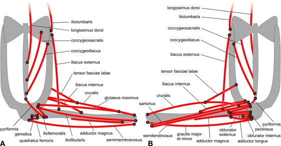
Comparative analysis of the anuran pelvic and thigh musculoskeletal system revealed that the thigh extensors, responsible for the initial phase of jump, the propulsive stroke in swimming and, if used asynchronously, also for walking, are least affected by the transformations observed between anurans and their temnospondyl ancestors (as reflected in contemporary caudates). The iliac shaft and urostyle, two of the most important anuran apomorphies, represent skeletal support for muscles that are mostly protractors of the femur or are important in attaining a crouching position, a necessary prerequisite for rapid escape. All of these muscles originate or insert on the iliac shaft. As the orientation of the pubis, ischium and ilium is the same in anurans, caudates and by inference also in their temnospondyl ancestors, it is probable that the pelvis was shifted from the sacral vertebra posteriorly along the reduced and stiffened tail (urostyle) by the elongation of the illiac shaft. Thus, the original vertical orientation of the ilium was maintained (which is also demonstrated by stable origins of the glutaeus maximus, iliofemoralis and iliofibularis on the tuber superius) and the shaft itself is a new structure. A review of functional analysis of anuran locomotion suggests some clear differences from that in caudates, suggesting that terrestrial jumping may have been a primary locomotor activity, from which other types of anuran locomotion are derived.


Figures

Fig. 1
Fig. 1
Diagrammatic representation of the pelvic and thigh muscles in (A) dorsal and (B) ventral views.
Fig. 2–1
Fig. 2–1
Discoglossus pictus. (A) Pelvic region (dorsal view), skin removed. (B) Same as in A but muscles attached to the urostyle prepared on the left side. (C) Surface layer of thigh muscles (ventral view). (D) Surface layer of thigh muscles (dorsal view). (E) Same as in D but glutaeus maximus partly removed. (F) Same as in C but gracilis major partly removed (ventral view). (G) Same as in D but glutaeus maximus and gracilis major removed (dorsal view). (H) Deep layer of thigh muscles (ventral view). (I) Same as in G but semimembranosus removed. Origin of glutaeus maximus marked by arrow. (J) Femur (ventral view), with insertions of obturator externus and adductor magnus. (K) The deepest layer (except for the cruralis) of thigh muscles (dorsal view). (L) Pelvis (dorsolateral view). (M) Same as in L but iliofemoralis removed. E is male and all others are female.
Fig. 2–2
Fig. 2–2
Discoglossus pictus. (A) Semidiagram of some pelvic and thigh muscles (dorsal view). (B) Origins of muscles on the right lateral surface of the pelvic girdle. (C) Origins of muscles on the inner surface of the right ilium. Based on skeleton DPFNSP 6514a (sex not recorded).
Fig. 3–1
Fig. 3–1
Ascaphus truei. (A) Pelvic region (dorsal view), skin removed. (B) Same as in A but right longissimus dorsi removed. (C) Pelvis (dorsolateral view). (D) Surface layer of thigh muscles (ventral view); arrow marks origin of sartorius-semitendinosus complex from obturator externus. (E) Surface layer of thigh muscles (dorsal view). (F) Same as in D but sartorius interrupted and partly raised, and gracilis-semimembranosus complex and cruralis partly removed. (G) Same as in E but glutaeus maximus partly removed. Arrow marks position of tendinous inscription of semimembranosus where caudalipuboischiotibialis is attached. (H) Same as in F but common insertion of sartorius-semitendinosus and gracilis-semimembranosus complexes cut and displaced to show their joint ligament. (I) Same as in G but iliofibularis and glutaeus maximus removed. (J) Same as in H but middle part of sartorius-semitendinosus removed and their proximal heads slightly raised to show their origins from obturator externus. (K) Middle layer of thigh muscles (dorsal view); cruralis and semimembranosus-gracilis complex partly displaced. (L) The deepest layer of thigh muscles (ventral view). (M) The deepest layer of thigh muscles (dorsal view); semimembranosus removed and distal part of caudalipuboischiotibialis detached. All illustrations represent male.
Fig. 3–2
Fig. 3–2
Ascaphus truei. (A) Semidiagram of some pelvic and thigh muscles (dorsal view). (B) Origins of muscles on right lateral surface of pelvic girdle. (C) Attachments of muscles on inner surface of right ilium. Based on skeleton DPFNSP 6538a (sex not recorded).
Fig. 4–1
Fig. 4–1
Barbourula busagensis. (A) Pelvic region (dorsal view), skin removed. Posterior margin of transversus marked by arrow. (B) Same as in A but fascia where transversus inserts partly removed to display longissimus dorsi-coccygeosacralis. (C) Same as in B but transversus on both sides detached from epaxial muscles to show both parts of iliacus externus. (D) Same as in C but in left dorsolateral view. (E) Surface layer of thigh muscles (ventral view); broken line marks origin of sartorius-semitendinosus on fascia of adductor magnus. (F) Surface layer of thigh muscles (dorsal view); posterior margin of transversus marked by arrow. (G) Same as in E but rectus abdominis and wall of abdominal cavity removed, posterior muscles partly isolated from each other. Arrow marks proximal margin of sartorius-semitendinosus. Inset illustrates outer portion of tensor fasciae latae. (H) Same as in F but fascia removed and muscles partly isolated from each other. Posterior margin of transversus marked by arrow. (I) Same as in G but dorsal head of semitendinosus partly removed. Arrow marks proximal margin of sartorius-semitendinosus. (J) Same as in H but glutaeus maximus partly removed to show its insertion. (K) Same as in I but gracilis major partly removed. Arrow marks proximal border of sartorius-semitendinosus. (L) Same as in J but glutaeus maximus removed. (M) Same as in K but sartorius-semitendinosus displaced and partly turned in order to expose ventral head of semitendinosus. Arrow marks tendon by which semitendinosus inserts in fascia separating both heads of adductor magnus. (N) Same as in L but iliofibularis removed. (O) Same as in M but sartorius-semitendinosus and semimembranosus removed. (P) Same as in N but gracilis major and minor removed. (Q) Same as in O but ventral head of adductor magnus detached and partly turned to show inner arrangement. (R) Same as in P but semimembranosus removed. (S) Same as in Q but adductor magnus removed. (T) Deep muscles of thigh (ventral view). (U) Deep muscles of thigh (dorsal view). (V) Deep proximal muscles of thigh (ventral view); tensor fasciae latae partly removed to show its origin.
Fig. 4–1
Fig. 4–1
Barbourula busagensis. (A) Pelvic region (dorsal view), skin removed. Posterior margin of transversus marked by arrow. (B) Same as in A but fascia where transversus inserts partly removed to display longissimus dorsi-coccygeosacralis. (C) Same as in B but transversus on both sides detached from epaxial muscles to show both parts of iliacus externus. (D) Same as in C but in left dorsolateral view. (E) Surface layer of thigh muscles (ventral view); broken line marks origin of sartorius-semitendinosus on fascia of adductor magnus. (F) Surface layer of thigh muscles (dorsal view); posterior margin of transversus marked by arrow. (G) Same as in E but rectus abdominis and wall of abdominal cavity removed, posterior muscles partly isolated from each other. Arrow marks proximal margin of sartorius-semitendinosus. Inset illustrates outer portion of tensor fasciae latae. (H) Same as in F but fascia removed and muscles partly isolated from each other. Posterior margin of transversus marked by arrow. (I) Same as in G but dorsal head of semitendinosus partly removed. Arrow marks proximal margin of sartorius-semitendinosus. (J) Same as in H but glutaeus maximus partly removed to show its insertion. (K) Same as in I but gracilis major partly removed. Arrow marks proximal border of sartorius-semitendinosus. (L) Same as in J but glutaeus maximus removed. (M) Same as in K but sartorius-semitendinosus displaced and partly turned in order to expose ventral head of semitendinosus. Arrow marks tendon by which semitendinosus inserts in fascia separating both heads of adductor magnus. (N) Same as in L but iliofibularis removed. (O) Same as in M but sartorius-semitendinosus and semimembranosus removed. (P) Same as in N but gracilis major and minor removed. (Q) Same as in O but ventral head of adductor magnus detached and partly turned to show inner arrangement. (R) Same as in P but semimembranosus removed. (S) Same as in Q but adductor magnus removed. (T) Deep muscles of thigh (ventral view). (U) Deep muscles of thigh (dorsal view). (V) Deep proximal muscles of thigh (ventral view); tensor fasciae latae partly removed to show its origin.
Fig. 4–2
Fig. 4–2
Barbourula busagensis. (A) Semidiagram of some pelvic and thigh muscles (dorsal view). (B) Origins of muscles on right lateral surface of pelvic girdle. (C) Insertions of muscles on inner surface of right ilium. Based on skeleton DPFNSP 6554a (female).
Fig. 5–1
Fig. 5–1
Bombina orientalis. (A) Pelvic region (dorsal view), skin removed. (B) Pelvis (dorsal view); muscles originating on right side of urostyle prepared, coccygeoiliacus removed. (C) Pelvis and proximal part of left femur with deep muscles (dorsolateral view). (D) Surface layer of thigh muscles (ventral view). (E) Surface layer of thigh muscles (dorsal view). (F) Same as in D but gracilis major removed, semimembranosus partly removed and glutaeus maximus partly displaced. (G) Same as in E but glutaeus maximus partly removed. (H) Same as in F but semitendinosus (caput dorsale) displaced to demonstrate its attachments. (I) Same as in G but semimembranosus removed and iliofibularis partly removed to show position of adductor magnus. (J) Same as in H but adductor magnus displaced and turned. (K) Same as in I but semimembranosus partly removed to show positions of gemellus and iliofemoralis. (L) Same as in J but all muscles inserting on femur, except for iliacus internus, removed. (M) Same as in K but all muscles except for iliacus externus and iliacus internus removed. A–C, K and M are male; D–J and L are female.
Fig. 5–2
Fig. 5–2
Bombina orientalis. (A) Semidiagram of some pelvic and thigh muscles (dorsal view). (B) Insertions of muscles on right lateral surface of pelvic girdle. (C) Insertions of muscles on inner surface of right ilium. Based on skeleton DPFNSP 6583 (male).
Fig. 6–1
Fig. 6–1
Pelobates fuscus. (A) Pelvis (dorsal view); most muscles originating on iliac shaft, except for coccygeoiliacus, removed. Note position and extent of sacral diapophyses. (B) Pelvis (dorsal view); superficial muscles around iliac joint removed. Note segmentation of longissimus dorsi. (C) Pelvis (left lateral view); muscles inserted on posterior part of iliac shaft removed. (D) Same as in C but some muscle insertions on posterior part of iliac shaft preserved, tensor fasciae latae partly removed. Sacral diapophysis and iliac shaft disarticulated. (E) Surface layer of thigh muscles (ventral view). (F) Surface layer of thigh muscles (dorsal view). Accessory head of plantaris longus marked by arrow. (G) Same as in E but gracilis major partly displaced to show origin of sartorius. (H) Same as in F but glutaeus maximus partly removed to show its insertion on cruralis, and two portions of cruralis. Plantaris longus removed. (I) Same as in G but sartorius and adductor magnus removed. (J) Same as in H but semimembranosus removed. (K) Deep layer of thigh muscles (ventral view). Pectineus partly displaced to demonstrate border with adductor longus. (L) Deep layer of thigh muscles (dorsal view). A, B–D, H and K are male; E–G, I, J and L are female.
Fig. 6–2
Fig. 6–2
Pelobates fuscus. (A) Semidiagram of some pelvic and thigh muscles (dorsal view). (B) Origins of muscles on right lateral surface of pelvic girdle. (C) Insertions of muscles on inner surface of right ilium. Based on skeleton DPFNSP 6333a (male).
Fig. 7–1
Fig. 7–1
Bufo guttatus. (A) Pelvis (dorsal view); skin removed. (B) Pelvis (dorsal view); right iliacus externus removed. (C) Pelvis (left dorsolateral view). (D) Pelvis (left dorsolateral view); only deep proximal muscles preserved. (E) Surface layer of thigh muscles in ventral view. The sartorius slightly raised to demonstrate its size. (F) Surface layer of thigh muscles (dorsal view). (G) Same as in E but sartorius partly removed (see arrow). (H) Same as in F but glutaeus maximus partly displaced to demonstrate extent of its fusion with cruralis. (I) Same as in G but gracilis major removed. (J) Same as in H but glutaeus maximus removed. (K) Deep proximal thigh muscles (ventral view). (L) Deep thigh muscles (dorsal view). All are female.
Fig. 7–2
Fig. 7–2
Bufo guttatus. (A) Semidiagram of some pelvic and thigh muscles (dorsal view). (B) Origins of muscles on right lateral surface of pelvic girdle. (C) Insertions of muscles on the inner surface of the right ilium. Based on skeleton DPFNSP 6582 (male).
Fig. 8–1
Fig. 8–1
Rana esculenta. (A) Pelvis (dorsal view); fascia removed on right side. (B) Pelvis (dorsal view); right coccygeoiliacus removed. (C) Pelvis and proximal part of left femur with deep muscles (lateral view). (D) Same as in C but iliacus internus removed. (E) Surface layer of thigh muscles (ventral view). (F) Surface layer of thigh muscles (dorsal view). (G) Same as in E but gracilis minor removed; gracilis major displaced to show ventral head of semitendinosus. (H) Same as in F but glutaeus maximus cut off proximally to demonstrate its insertion on cruralis. Note a. circumflexa femoris lateralis between iliacus internus and cruralis. (I) Same as in G but distal section of sartorius raised to show its attachment to caput tibiae. (J) Same as in H but semimembranosus displaced to show deep layer of dorsal thigh muscles. (K) Same as in I but sartorius removed. (L) Same as in J but semimembranosus removed. (M) Same as in K but gracilis major removed. (N) Same as in L but adductor magnus partly removed. A and B are female; C–N are male.
Fig. 8–2
Fig. 8–2
Rana esculenta. (A) Semidiagram of some pelvic and thigh muscles (dorsal view). (B) Origins of muscles on right lateral surface of pelvic girdle. (C) Insertions of muscles on inner surface of right ilium. Based on skeleton DPFNSP 6298b (female).
Fig. 9–1
Fig. 9–1
Xenopus laevis. (A) Pelvis (dorsal view); skin removed. Border between iliacus externus and longissimus dorsi marked by arrow. (B) Pelvis (dorsal view); right iliacus externus partly displaced to demonstrate its layers. (C) Pelvis (dorsal view); right iliacus externus removed. (D) Surface layer of thigh muscles (ventral view). (E) Surface layer of thigh muscles (dorsal view). (F) Same as in D but surface fascia and gracilis minor removed. Note extent of rectus abdominis. (G) Same as in E but glutaeus maximus partly displaced and semimembranosus removed. (H) Same as in F but sartorius removed and semitendinosus partly displaced to show its two parts. (I) Same as in G but glutaeus maximus and iliofibularis removed, and cruralis partly displaced to demonstrate extent of adductor magnus. (J) Deep thigh muscles (ventral view). (K) Insertion of iliacus externus on femur (dorsal view). All are females.
Fig. 9–2
Fig. 9–2
Xenopus laevis. (A) Semidiagram of some pelvic and thigh muscles (dorsal view). (B) Origins of muscles on right lateral surface of pelvic girdle. (C) Insertions of muscles on inner surface of right ilium. Based on skeleton DPFNSP 6411a (sex not recorded).
Fig. 10–1
Fig. 10–1
Pipa pipa. (A) Pelvis (dorsal view); skin removed. (B) Right part of pelvis (dorsal view); longissimus dorsi and iliacus externus removed. Note structure of iliosacral joint. (C) Pelvis (left dorsolateral view). Note extent of longissimus dorsi and division of iliacus externus. (D) Pelvis (left lateral view). Pulmonum proprius removed, its insertion marked by arrow. (E) Surface layer of thigh muscles (ventral view). Posterior part of rectus abdominis raised to show its insertion between cruralis and sartorius. (F) Surface layer of thigh muscles (dorsal view). (G) Same as in E but rectus abdominis flapped posteriorly to show adductor longus. (H) Same as in F but longissimus dorsi partly removed to show three parts of iliacus externus. (I) Same as in G but rectus abdominis flapped anteriorly to show partly displaced semitendinosus. (J) Same as in H but glutaeus maximus removed and semitendinosus partly displaced. (K) Deep thigh muscles (ventral view). (L) Insertion of three parts of iliacus externus on femur (dorsal view).
Fig. 10–2
Fig. 10–2
Pipa pipa. (A) Semidiagram of some pelvic and thigh muscles (dorsal view). Internal part of iliacus externus not illustrated. (B) Origins of muscles on right lateral surface of pelvic girdle. (C) Insertions of muscles on inner surface of right ilium. All images represent female. Based on skeleton DPFNSP 6385a (male).
Fig. 15
Fig. 15
Diagrammatic representation of some pelvic and thigh muscles in caudates (A) and anurans (B), in dorsal view.
Fig. 12
Fig. 12
Thigh muscles in Necturus maculosus. (A–G) Ventral view; (H–K) dorsal view. (A) Superficial level of thigh muscles (ventral view). (B) Same as in A but caudalipuboischiotibialis and ischiocaudalis exposed. (C) Same as in B but puboischiotibialis slightly raised to demonstrate its extent. (D) Same as in C but puboischiotibialis removed. (E) Same as in D but ischiofemoralis partly removed. (F) Same as in E but anterior portion of puboischiofemoralis externus removed so inner surface of pubis is exposed. (G) Surface level of thigh muscles (dorsal view). (H) Same as in G but ischioflexorius (and puboischiotibialis from the ventral side) removed. Posterior part of the iliotibialis slightly raised to demonstrate its posterior margin. (I) Same as in H but posterior part of iliotibialis removed. (J) Same as in I but iliofibularis removed. (K) Same as in J but anterior part of iliotibialis removed to demonstrate deep layer of muscles attached to femur. In A–C, the tendinous inscription in the puboischiotibialis is marked by arrows.
Fig. 14
Fig. 14
Thigh muscles in adult Salamandra. (A–F) Ventral view; (G–K) dorsal view. (A) Surface layer of thigh muscles (ventral view). (B) Same as in A but caudalipuboischiotibialis and ischiocaudalis exposed. (C) Same as in B but ischioflexorius exposed. (D) Same as in C but puboischiotibialis detached proximally. (E) Same as in D but puboischiotibialis removed. (F) Same as in E but puboischiofemoralis removed to expose pelvis and proximal section of femur. (G) Surface layer of thigh muscles (posterodorsal view). (H) Same as in G but dorsal view. (I) Same as in G but hypaxial trunk muscles partly removed. (J) Same as in I but iliotibialis removed to show deep layer. (K) Same as in J but most anterior of deep muscles removed.
Fig. 16
Fig. 16
Origins of some muscles in anuran (A) and caudate (B) pelvis, seen in right lateral view.
Fig. 13
Fig. 13
Thigh muscles in larval Ambystoma. (A–F) Ventral view; (G–J) dorsal view; (K–L) right lateral view. (A) Surface layer of thigh muscles (ventral view). Tendinous inscription in puboischiotibialis marked by arrow. (B) Same as in A but caudalipuboischiotibialis and ischiocaudalis exposed. Tendinous inscription in puboischiotibialis marked by arrow. (C) Same as in B but puboischiotibialis detached proximally to show anterior attachment of ischiocaudalis. (D) Same as in C but puboischiotibialis removed. (E) Same as in D but puboischiotibialis removed to show ischiofemoralis. In E1pubotibialis is partly removed to show pubofemoralis (marked by arrow). (F) Deep muscles of thigh (ventral view). (G) Superficial level of thigh muscles (dorsal view). (H) Same as in G but dorsal trunk muscles removed to expose ilium. (I) Same as in H but posterior muscles detached proximally and partly displaced. (J) Same as in I but posterior muscles removed to show pattern of deep muscles. (K) Proximal attachment of thigh muscles (lateral view). (L) Same as in K but posterior muscles removed. Muscles in A–C slightly detached from each other.
Fig. 11
Fig. 11
Pelobates fuscus. Extreme positions of the pelvis during burrowing.

Similar articles

Cited by

References

    1. Anderson JS, Reisz RR, Scott D, Fröbisch NB, Sumida SS. A stem batrachian from the early Permian of Texas and the origin of frogs and salamanders. Nature. 2008;453:515–518. - PubMed
    1. Blanco MJ, Sanchiz B. Evolutionary mechanisms of rib loss in anurans: a comparative developmental approach. J Morphol. 2000;244:57–67. - PubMed
    1. Böker H. Einführung in Die Vergleichende Biologische Anatomie der Wirbeltiere. Vol. 1. Jena: Gustav Fischer; 1935.
    1. Boy JA. Die Larven der rhachitomen Amphibien (Amphibia: temnospondyli; Karbon-Trias) Paläont Z. 1974;48:236–268.
    1. Boy JA, Sues H-D. Branchiosaurs: larvae, metamorphosis and heterochrony in temnospondyls and seymouriamorphs. In: Heatwole H, Carroll R, editors. Amphibian Biology – Paleontology. Chipping Norton: Surrey Beatty & Sons; 2000. pp. 1150–1197.

Publication types