Comparative Study
. 2009 Jun;33(6):747-60.
doi: 10.1016/j.dci.2008.12.007. Epub 2009 Jan 21.

Identification of a porcine DC-SIGN-related C-type lectin, porcine CLEC4G (LSECtin), and its order of intron removal during splicing: comparative genomic analyses of the cluster of genes CD23/CLEC4G/DC-SIGN among mammalian species

Affiliations
Comparative Study

Identification of a porcine DC-SIGN-related C-type lectin, porcine CLEC4G (LSECtin), and its order of intron removal during splicing: comparative genomic analyses of the cluster of genes CD23/CLEC4G/DC-SIGN among mammalian species

Y W Huang et al. Dev Comp Immunol. 2009 Jun.

Abstract

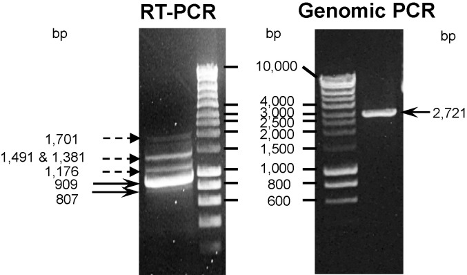
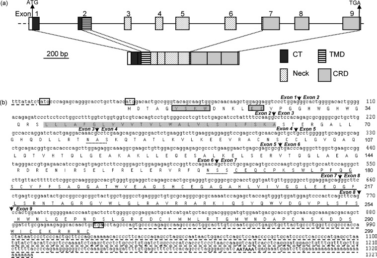
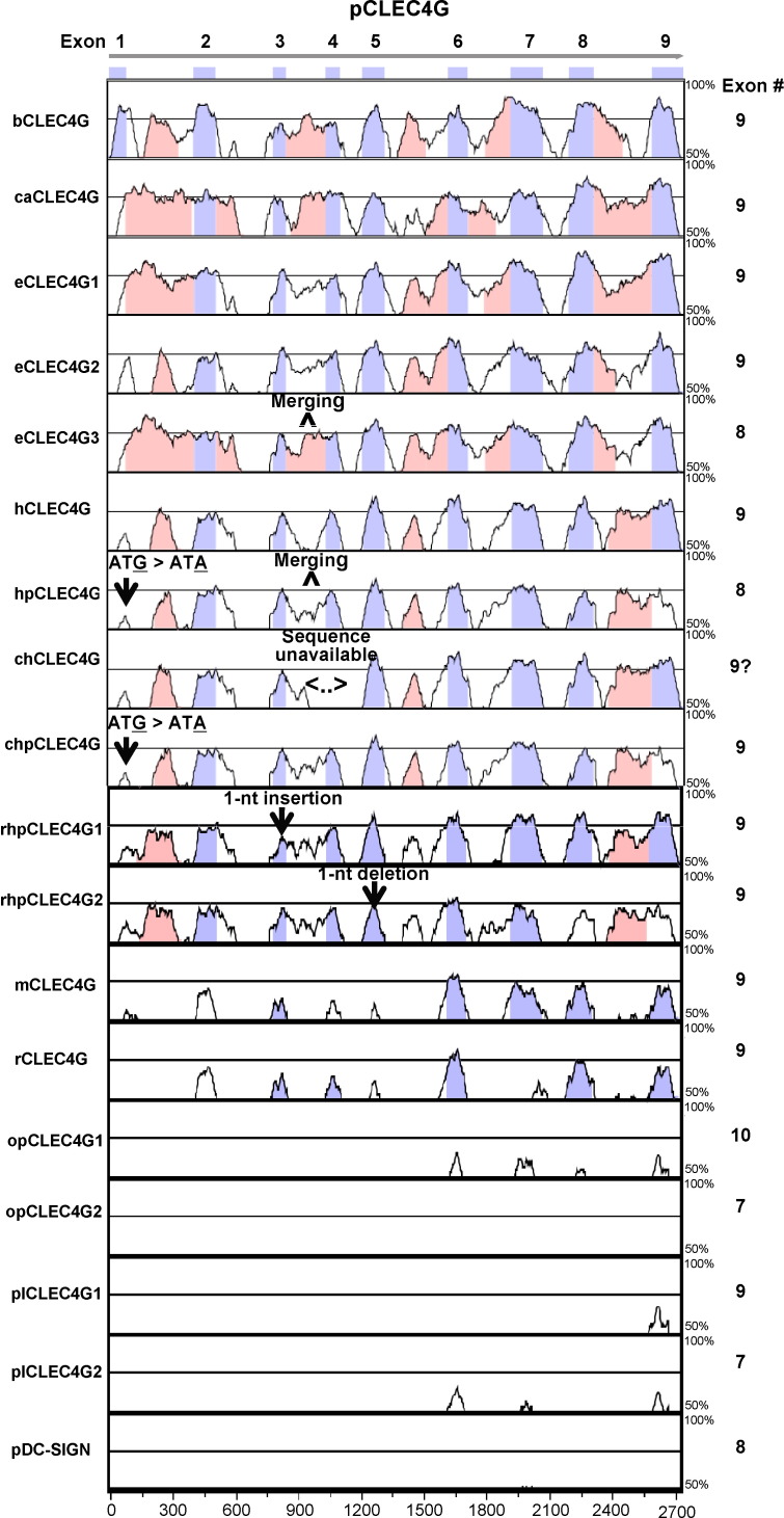
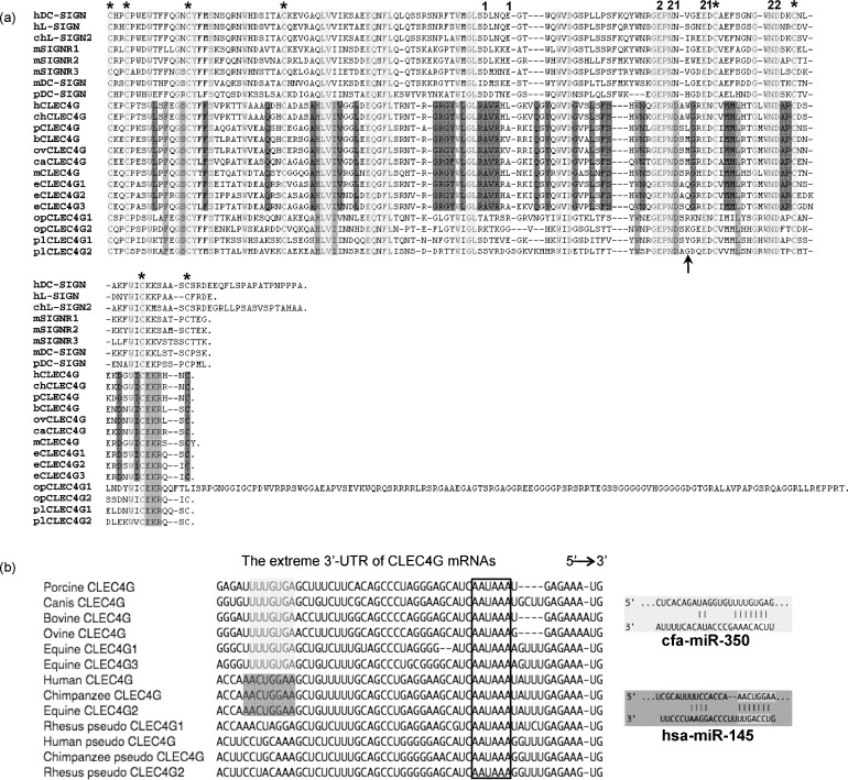
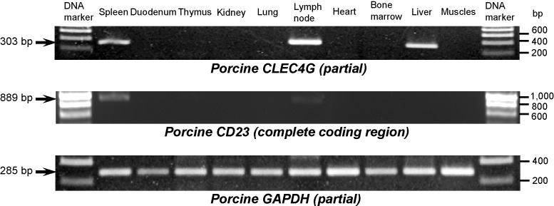
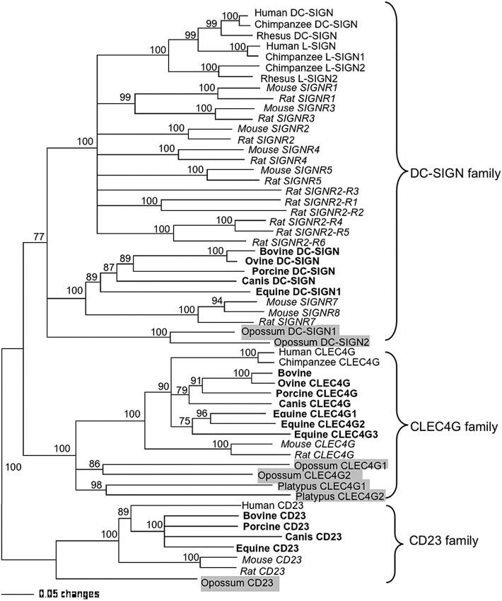
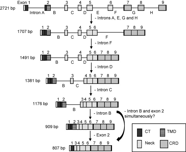
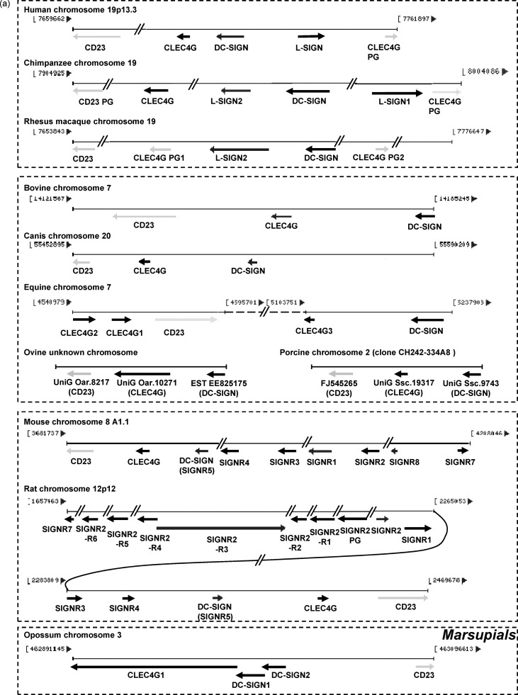
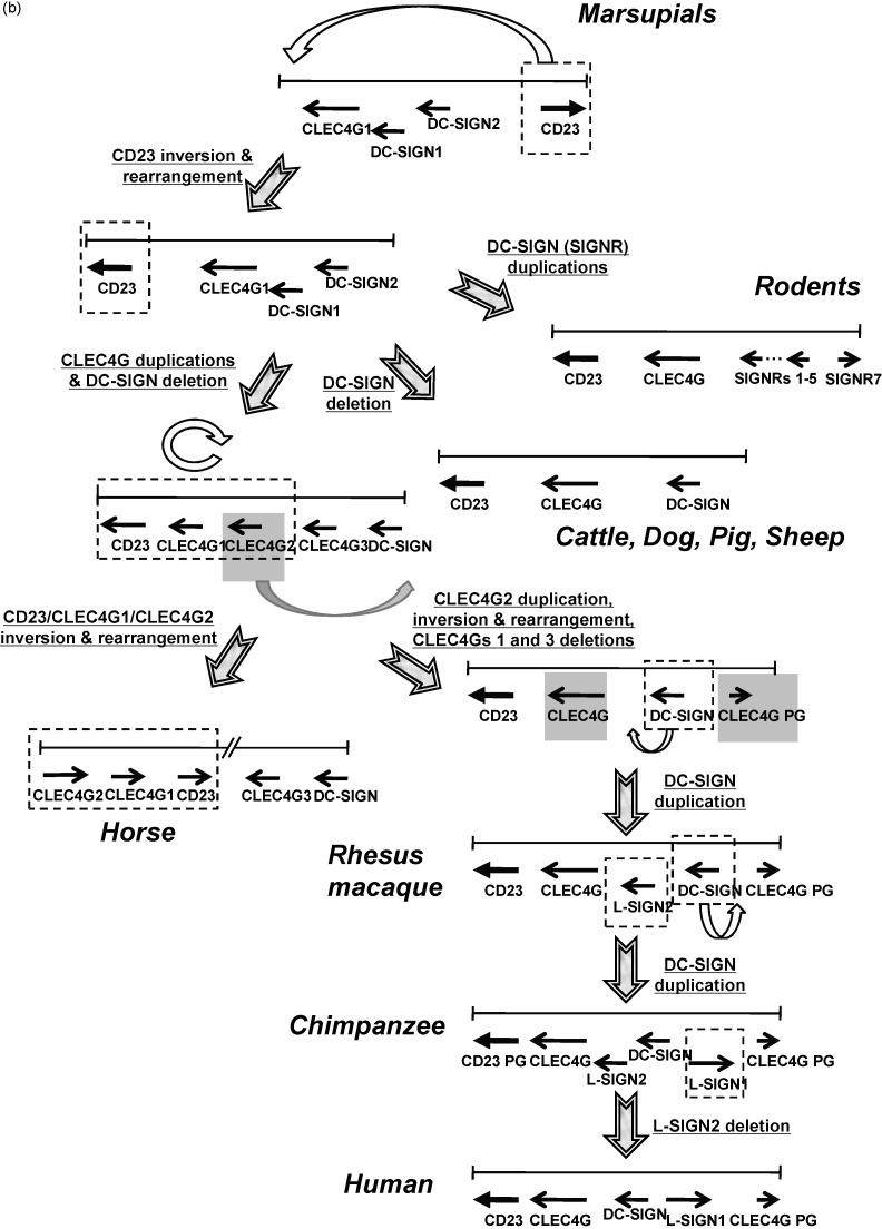
Human CLEC4G (previously named LSECtin), DC-SIGN, and L-SIGN are three important C-type lectins capable of mediating viral and bacterial pathogen recognitions. These three genes, together with CD23, form a lectin gene cluster at chromosome 19p13.3. In this study, we have experimentally identified the cDNA and the gene encoding porcine CLEC4G (pCLEC4G). Full-length pCLEC4G cDNA encodes a type II transmembrane protein of 290 amino acids. pCLEC4G gene has the same gene structure as the human and the predicted bovine, canis, mouse and rat CLEC4G genes with nine exons. A multi-species-conserved site at the extreme 3'-untranslated region of CLEC4G mRNAs was predicted to be targeted by microRNA miR-350 in domesticated animals and by miR-145 in primates, respectively. We detected pCLEC4G mRNA expression in liver, lymph node and spleen tissues. We also identified a series of sequential intermediate products of pCLEC4G pre-mRNA during splicing from pig liver. The previously unidentified porcine CD23 cDNA containing the complete coding region was subsequently cloned and found to express in spleen, thymus and lymph node. Furthermore, we compared the chromosomal regions syntenic to the human cluster of genes CD23/CLEC4G/DC-SIGN/L-SIGN in representative mammalian species including primates, domesticated animal, rodents and opossum. The L-SIGN homologues do not exist in non-primates mammals. The evolutionary processes of the gene cluster, from marsupials to primates, were proposed based upon their genomic structures and phylogenetic relationships.


Figures

Fig. 1
Fig. 1
Amplification of the intermediate and mature products (isoforms) of pCLEC4G pre-mRNA during splicing from pig liver by RT-PCR and amplification of pCLEC4G gene from pig genomic DNA by genomic PCR. Dashed-line arrows showed the spliced intermediate products and solid-line arrows indicated the isoforms and pCLEC4G gene.
Fig. 2
Fig. 2
(a) Gene structure of pCLEC4G gene. The upper row displayed the exon allocation of domains. The lower row represented the domain structure of the putative pCLEC4G coding region. CT: cytoplasmic tail; TMD: transmembrane domain; CRD: carbohydrate recognition domain. Un-translated regions in exons 1 and 9 were shown as open boxes. (b) Complete nucleotide sequence of pCLEC4G cDNA and its deduced amino acid sequence. Extra nucleotide sequences at both termini in the non-coding region of pCLEC4G cDNA that were not determined in this study were included with dashed underlines (available from nucleotide sequence with GenBank accession number AK232603). The two in-frame initiation codons and the stop codon were boxed. Two potential internalization motifs, YSKW and EE in the CT, were indicated by gray boxes. The putative TMD was indicated by a gray background and the carbohydrate recognition domain (CRD) is underlined. Two predicted glycosylation sites in the neck region were indicated by dotted underlines. The polyadenylation signal (AATAAA) was indicated by capitals. Arrows show the boundary of exons.
Fig. 3
Fig. 3
Comparison of the gene sequences and numbers of exons of pCLEC4G gene with other CLEC4G homologues as well as with pDC-SIGN gene generated by the mVISTA program. Conserved regions between pairs of sequences are displayed as peaks of similarity (Y axis) relative to the positions of the gene sequence of pCLEC4G (X axis). The blue-violet boxes above the plots represent the nine exons of the pCLEC4G gene. The peaks in the same color indicate conserved regions within exons while the peaks in pink color denote conserved regions within introns. The cutoff value of percent identity is set to 70%. The human and chimpanzee CLEC4G pseudogenes lost their protein-coding ability due to a point mutation (G to A) at the proposed start codon. The two rhesus macaque CLEC4G pseudogenes are unable to encode functional CLEC4G proteins due to a 1-nt insertion or a 1-nt deletion leading to the frame shift. The exon 4 sequence of chimpanzee CLEC4G gene is not available thus far. Abbreviations: porcine CLEC4G (pCLEC4G), bovine CLEC4G (bCLEC4G), canis CLEC4G (caCLEC4G), equine CLEC4G (eCLEC4G), human CLEC4G (hCLEC4G), human CLEC4G pseudogene (hpCLEC4G), chimpanzee CLEC4G (chCLEC4G), chimpanzee CLEC4G pseudogene (chpCLEC4G), rhesus macaque CLEC4G pseudogene (rhpCLEC4G), mouse CLEC4G (mCLEC4G), rat CLEC4G (rCLEC4G), opossum CLEC4G (opCLEC4G), platypus CLEC4G (plCLEC4G), and porcine DC-SIGN (pDC-SIGN).(For interpretation of the references to color in this figure legend, the reader is referred to the web version of the article.)
Fig. 4
Fig. 4
(a) Alignment of amino acid sequences of the CRD of pCLEC4G and other CLEC4G as well as DC-SIGN homologues among various vertebrate species. Amino acid residues that form Ca2+-binding site 1 are indicated by “1”, residues that form Ca2+-binding site 2 are indicated by “2”, and conserved cysteine residues involved in disulfide bond formations are indicated by “*”. An arrow indicated the aa position 265 following the EPN motif where Trp residue of hCLEC4G had been proposed to interact with the GlcNAc residue of a disaccharide GlcNAcβ1-2Man. Alignment gaps are indicated by dashes. The consensus residues with identical amino acids among all placental CLEC4G members, all mammalian CLEC4G members, and both DC-SIGN and CLEC4G members are highlighted in dark gray, middle gray and light gray, respectively. Abbreviation: ovine CLEC4G (ovCLEC4G). Other abbreviations are the same as those in Fig. 3. (b) Prediction of a multi-species-conserved microRNA (miRNA) target sequence at the three prime untranslated regions (3′-UTR) of domesticated animal and primate CLEC4G mRNAs by the TargetScan program. Only one common region (nt position 1247–1254 corresponding to pCLEC4G, see Fig. 2b) that was targeted by miR-350 in domesticated animal CLEC4G members except equine CLEC4G2 (highlighted in light gray) and by miR-145 in human and chimpanzee CLEC4Gs as well as equine CLEC4G2 (highlighted in dark gray), respectively, was identified at the extreme 3′UTR. The polyadenylation signals (AAUAAA) were indicated by boxed. The respective predicted pairing of target region and miRNA was shown on the right.
Fig. 5
Fig. 5
Detection of pCLEC4G and pCD23 mRNA expression in selected pig tissues by RT-PCR. Pig tissue cDNAs were used as templates in PCR reactions with primers PLST-E67F/PLST-E89R, PCD23-F/PCD23-R and porcine GAPDH-specific primers, respectively. Arrows indicate the sizes of expected PCR products.
Fig. 6
Fig. 6
Phylogenetic tree constructed by the neighbor-joining method based upon the amino acid sequences of DC-SIGN family, CLEC4G family and CD23 family proteins. Bootstrap values are indicated for each node from 1000 re-sampling. Proteins from rodents (mouse and rat), domesticated animals and non-placentals (opossum and platypus) were highlighted in italic, bold and highlighted gray, respectively.
Fig. 7
Fig. 7
Proposed order of intron removal from porcine CLEC4G pre-mRNA. Boxes with numbers 1–9 represent the nine pCLEC4G exon sequences. The eight intron sequences, letters A to H, are indicated by the black lines between the exons. The arrows show the splicing pathway. The gene, splicing intermediate products and isoforms detected by RT-PCR, are indicated with their respective sizes shown on the left.
Fig. 8
Fig. 8
(a) Schematic representations of the clusters of genes CD23/CLEC4G/DC-SIGN in human, chimpanzee, rhesus macaque, bovine, canis, equine, mouse, rat and opossum. The relative size and orientation of CLEC4G, DC-SIGN, L-SIGN and SIGNR genes on their respective chromosome were shown as black arrows, whereas the CD23 and pseudogenes (PG) were indicted as light gray arrows. The genomic loci and coordinates were described in Section 2. (b) Proposed evolutionary processes of the cluster of genes CD23/CLEC4G/DC-SIGN from marsupials to primates based on their genomic structures and phylogenetic relationships. Only the orientations and arrangement of the genes were shown as black arrows, whereas the relative size of the genes and genomic coordinates were not shown. Notched arrows indicated the proposed key processes. Possible gene mutation events including inversion, rearrangement, duplication and deletion in each process were described with underlines adjacent to the corresponding notched arrow.
Fig. 8
Fig. 8
(a) Schematic representations of the clusters of genes CD23/CLEC4G/DC-SIGN in human, chimpanzee, rhesus macaque, bovine, canis, equine, mouse, rat and opossum. The relative size and orientation of CLEC4G, DC-SIGN, L-SIGN and SIGNR genes on their respective chromosome were shown as black arrows, whereas the CD23 and pseudogenes (PG) were indicted as light gray arrows. The genomic loci and coordinates were described in Section 2. (b) Proposed evolutionary processes of the cluster of genes CD23/CLEC4G/DC-SIGN from marsupials to primates based on their genomic structures and phylogenetic relationships. Only the orientations and arrangement of the genes were shown as black arrows, whereas the relative size of the genes and genomic coordinates were not shown. Notched arrows indicated the proposed key processes. Possible gene mutation events including inversion, rearrangement, duplication and deletion in each process were described with underlines adjacent to the corresponding notched arrow.

Similar articles

Cited by

References

    1. Cambi A., Koopman M., Figdor C.G. How C-type lectins detect pathogens. Cell Microbiol. 2005;7(4):481–488. - PubMed
    1. Weis W.I., Taylor M.E., Drickamer K. The C-type lectin superfamily in the immune system. Immunol Rev. 1998;163:19–34. - PubMed
    1. Conrad D.H., Ford J.W., Sturgill J.L., Gibb D.R. CD23: an overlooked regulator of allergic disease. Curr Allergy Asthma Rep. 2007;7(5):331–337. - PubMed
    1. Geijtenbeek T.B., Krooshoop D.J., Bleijs D.A. DC-SIGN–ICAM-2 interaction mediates dendritic cell trafficking. Nat Immunol. 2000;1(4):353–357. - PubMed
    1. Geijtenbeek T.B., Torensma R., van Vliet S.J., van Duijnhoven G.C., Adema G.J., van Kooyk Y., Figdor C.G. Identification of DC-SIGN, a novel dendritic cell-specific ICAM-3 receptor that supports primary immune responses. Cell. 2000;100(5):575–585. - PubMed

Publication types

MeSH terms

Associated data