Review
. 2009 Feb 27;61(2):86-100.
doi: 10.1016/j.addr.2008.09.012. Epub 2009 Jan 3.

Micro- and macrorheology of mucus

Affiliations
Review

Micro- and macrorheology of mucus

Samuel K Lai et al. Adv Drug Deliv Rev. .

Abstract

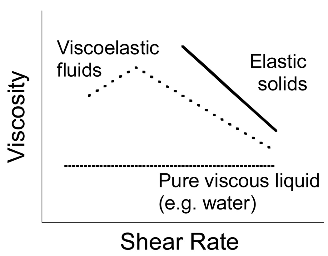
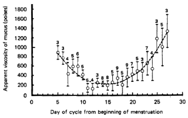
Mucus is a complex biological material that lubricates and protects the human lungs, gastrointestinal (GI) tract, vagina, eyes, and other moist mucosal surfaces. Mucus serves as a physical barrier against foreign particles, including toxins, pathogens, and environmental ultrafine particles, while allowing rapid passage of selected gases, ions, nutrients, and many proteins. Its selective barrier properties are precisely regulated at the biochemical level across vastly different length scales. At the macroscale, mucus behaves as a non-Newtonian gel, distinguished from classical solids and liquids by its response to shear rate and shear stress, while, at the nanoscale, it behaves as a low viscosity fluid. Advances in the rheological characterization of mucus from the macroscopic to nanoscopic levels have contributed critical understanding to mucus physiology, disease pathology, and the development of drug delivery systems designed for use at mucosal surfaces. This article reviews the biochemistry that governs mucus rheology, the macro- and microrheology of human and laboratory animal mucus, rheological techniques applied to mucus, and the importance of an improved understanding of the physical properties of mucus to advancing the field of drug and gene delivery.


Figures

Figure 1
Figure 1
Illustration of the steady state viscosity vs. shear rate profiles of liquids, solids, and viscoelastic substances. The viscosity of a liquid is constant, while the viscosity of a yielding solid decreases with time. However, the viscosity of a viscoelastic material is more complex. In the above example of a thixotropic fluid, the steady state viscosity first increases at low shear rates (shear thickening), then progressively decreases at larger shear rates (shear thinning).
Figure 2
Figure 2
Major biochemical features of gel-forming mucins. (A) Several mucin monomers are shown linked together in an oligomeric gel. (B) Mucin monomers are crosslinked end-to-end via disulfide bonds between disulfide-rich domains (labeled “D”) near the amino- and carboxyl-termini [45, 196, 197]. (C) Interspersed along each fiber are “naked” globular protein regions, with small exposed hydrophobic patches [198]. These regions are stabilized by multiple disulfide bonds. (D) Individual mucin fibers are densely glycosylated with O- and N-linked glycans, most of which are negatively charged with sialic acids or sulfate groups [45]. Figure is obtained from [6].
Figure 3
Figure 3
(A) Schematic representation of the length scale dependence of viscosity in a nanoscopically heterogeneous fluid. Non-adhesive particles that are significantly smaller than the mesh spacing undergo Brownian diffusion and probe the microscopic rheology. As the particle size approaches the dimensions of the mesh spacing, particle movement becomes hindered by the mesh microstructure at short time scales, leading to a mesophase rheology regime. Particles that are significantly larger than the mesh spacing probe the bulk or macroscopic rheology of the gel. (B) Schematic comparison of non-mucoadhesive rheological nanoprobes and conventional polymeric particles. Conventional nanoprobes are immobilized to mucin fibers via adhesive interactions. Their strongly hindered motion, as reflected by the small dimensions of the traces, suggest a markedly higher viscoelastic environment than the true local viscoelasticity of mucus. In contrast, the motion of non-mucoadhesive nanoprobes correctly reflects the local viscous and elastic contributions from the mucus mesh architecture.
Figure 4
Figure 4
Viscosity of various types of human mucus. (A) Dynamic oscillatory viscosity as a function of shear frequency for cervical mucus (red, [37, 138, 201]), nasal mucus (blue, [–93]), and lung mucus (black, [75, 78, 114, 199, 200]). Solid lines correspond to normal mucus, while dashed lines indicate mucus under disease conditions. The shaded region represents the range of values for all mucus types shown. (B) Steady shear viscosity as a function of shear rate for (a, b) chronic bronchitis mucus (green circle [202] and square [11]), (c) non-ovulatory cervical mucus (red, [70]), (d) normal gastric mucus (black line, [105]), (e) duodenal ulcer mucus (black dashed line, [105]), and (f) tears (blue, [111]). Thin dashed lines indicate the typical range of viscosity values for human mucus suggested by [6]. The thin solid line represents the viscosity of water (10−3 Pa–s).
Figure 5
Figure 5
Macrorheology of human cystic fibrosis sputum. (A) The frequency dependent elastic, G’(ω), and viscous moduli, G”(ω), of CF samples (n=6) were recorded at a constant strain amplitude of 1%. (B) Strain-dependent elastic, G’(ω), and viscous moduli, G”(ω), from 0.1–100% strain amplitude. (C) The steady state viscosity of CF sputum at shear rates between 10− 2−102 rad/s. Physiological rates in the normal lung are represented by the dotted line. Inset: viscosities of individual CF sputum samples at physiological shear rates. Figure obtained from ref [14].
Figure 6
Figure 6
Variations in the viscosity of cervical mucus from healthy, non-pregnant subjects with day in menstruation cycle (119 samples). The dotted line is drawn to emphasize the main feature of the graph. The number above each point indicates the number of samples averaged. Each vertical line indicates ± one standard deviation of individual readings about the mean. Figure obtained from [108].
Figure 7
Figure 7
Confocal micrograph of CF sputum showing DNA polymers in green (Yoyo-1 stain) and minimal mucin staining in red (UAE-Texas red). Figure obtained from [81].

References

    1. Knowles MR, Boucher RC. Mucus clearance as a primary innate defense mechanism for mammalian airways. J Clin Invest. 2002;109:571–577. - PMC - PubMed
    1. Chilvers MA, O'Callaghan C. Local mucociliary defence mechanisms. Paediatr Respir Rev. 2000;1:27–34. - PubMed
    1. McAuley JL, Linden SK, Png CW, King RM, Pennington HL, Gendler SJ, Florin TH, Hill GR, Korolik V, McGuckin MA. MUC1 cell surface mucin is a critical element of the mucosal barrier to infection. J Clin Invest. 2007;117:2313–2324. - PMC - PubMed
    1. Strombeck DR, Harrold D. Binding of cholera toxin to mucins and inhibition by gastric mucin. Infect Immun. 1974;10:1266–1272. - PMC - PubMed
    1. Kreyling WG, Semmler M, Moller W. Dosimetry and toxicology of ultrafine particles. J Aerosol Med. 2004;17:140–152. - PubMed

Publication types