. 2009 Jan;96(2):L22-4.
doi: 10.1016/j.bpj.2008.11.002.

Light-induced dark states of organic fluochromes enable 30 nm resolution imaging in standard media

Light-induced dark states of organic fluochromes enable 30 nm resolution imaging in standard media

David Baddeley et al. Biophys J. 2009 Jan.

Abstract

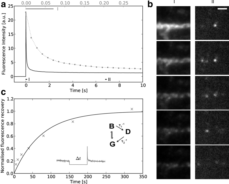
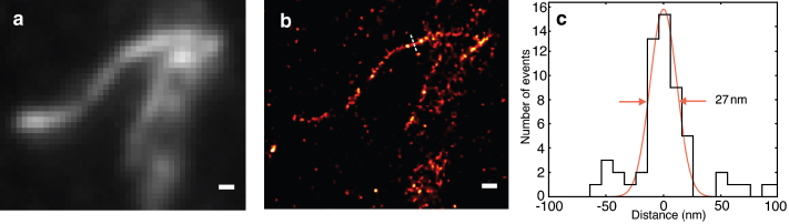
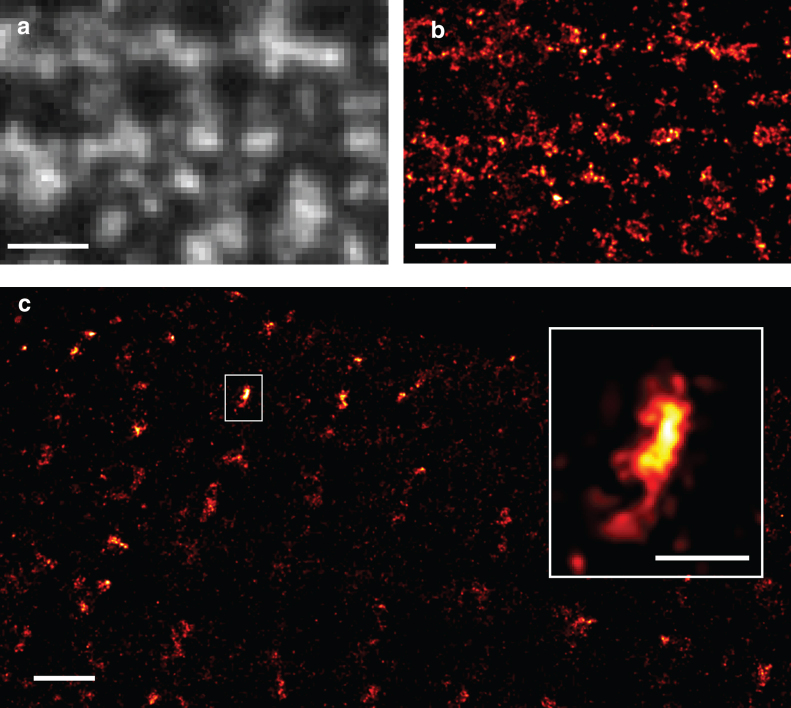
We show that high quantum efficiency fluorophores can exhibit reversible photobleaching. This observation provides the basis for an imaging technique we call reversible photobleaching microscopy. We demonstrate applicability of this technique using antibody labeled biological samples in standard aqueous (or glycerol based) media to produce far-field images at approximately 30 nm resolution. Our novel method relies on intense illumination to reversibly induce a very long-lived (>10 s) dark state from which single fluorochromes slowly return stochastically. As in other localization microscopy methods, reversible photobleaching microscopy localizes single fluorochromes, but has the advantage that specialized photoactivatible and photoswitchable molecules or special immersion/embedding media are not required.


Figures

Figure 1
Figure 1
Reversible photobleaching of Alexa 488. (a) Upon exposure to intense light a rapid decline in fluorescence of Alexa 488 is observed, followed by a plateau phase. The dotted line shows the initial decay on a magnified timescale (top axis). (b) Consecutive frames taken during initial bleaching (I), show the whole stained structure, whereas those taken during the plateau phase (II) show the stochastic reappearance of single fluorophores as they revert from the dark state. (c) After a period of darkness a significant recovery in the Alexa 488 fluorescence is observed with an approximately logarithmic dependence on the length of the dark period, implying a half time of ∼1 min. A simple scheme illustrates the dark state behavior, see text. Scale bar, 1 μm.
Figure 2
Figure 2
Imaging of Alexa 488 phalloidin labeled F-actin. (a) Diffraction limited image obtained by averaging the first 100 frames. (b) Image reconstructed from single fluorophore localization events, showing considerably better resolution. (c) Line profile along dotted line in b with a fitted Gaussian width of 27 nm. Scale bars, 200 nm.
Figure 3
Figure 3
RPM images of cardiac caveolin and RyRs. (a) TIRF image of caveolin-3 in the surface membrane of a rat ventricular myocyte (gray), (b) a RPM image of the same region—some of the smaller clusters are likely associated with single caveolae. (c) Clusters of RyRs at the membrane. The magnified cluster (inset) has a size of ∼60 × 180 nm corresponding to ∼12 of the large, ∼30 nm diameter receptors. Scale bars: a, 1 μm; b, 200 nm; and c, 1 μm; inset, 200 nm.

References

    1. Abbe E. Beiträge zur Theorie des Mikroskops und der mikroskopischen Wahrnehmung. Arch. f. mikroskop. Anat. 1873;9:413–420.
    1. Gustafsson M.G. Nonlinear structured-illumination microscopy: wide-field fluorescence imaging with theoretically unlimited resolution. Proc. Natl. Acad. Sci. USA. 2005;102:13081–13086. - PMC - PubMed
    1. Dyba M., Jakobs S., Hell S.W. Immunofluorescence stimulated emission depletion microscopy. Nat. Biotechnol. 2003;21:1303–1304. - PubMed
    1. Hofmann M., Eggeling C., Jakobs S., Hell S.W. Breaking the diffraction barrier in fluorescence microscopy at low light intensities by using reversibly photoswitchable proteins. Proc. Natl. Acad. Sci. USA. 2005;102:17565–17569. - PMC - PubMed
    1. Betzig E., Patterson G.H., Sougrat R., Lindwasser O.W., Olenych S., et al. Imaging intracellular fluorescent proteins at nanometer resolution. Science. 2006;313:1642–1645. - PubMed

Publication types