. 2009 Jan;96(2):566-82.
doi: 10.1016/j.bpj.2008.10.020.

Single-molecule fluorescence studies of a PH domain: new insights into the membrane docking reaction

Affiliations

Single-molecule fluorescence studies of a PH domain: new insights into the membrane docking reaction

Jefferson D Knight et al. Biophys J. 2009 Jan.

Abstract

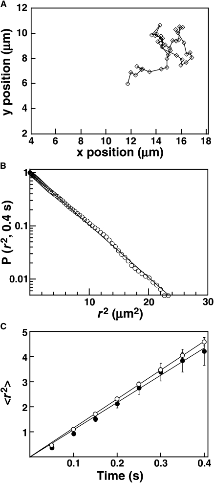
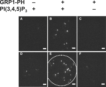
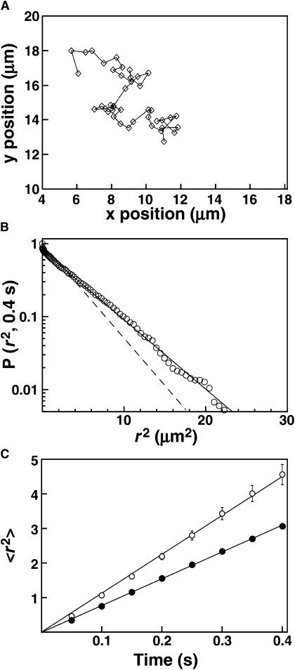
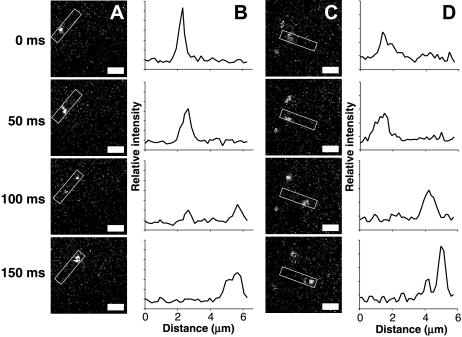
Proteins containing membrane targeting domains play essential roles in many cellular signaling pathways. However, important features of the membrane-bound state are invisible to bulk methods, thereby hindering mechanistic analysis of membrane targeting reactions. Here we use total internal reflection fluorescence microscopy (TIRFM), combined with single particle tracking, to probe the membrane docking mechanism of a representative pleckstrin homology (PH) domain isolated from the general receptor for phosphoinositides, isoform 1 (GRP1). The findings show three previously undescribed features of GRP1 PH domain docking to membranes containing its rare target lipid, phosphatidylinositol (3,4,5)-trisphosphate [PI(3,4,5)P(3)]. First, analysis of surface diffusion kinetics on supported lipid bilayers shows that in the absence of other anionic lipids, the PI(3,4,5)P(3)-bound protein exhibits the same diffusion constant as a single lipid molecule. Second, the binding of the anionic lipid phosphatidylserine to a previously unidentified secondary binding site slows both diffusion and dissociation kinetics. Third, TIRFM enables direct observation of rare events in which dissociation from the membrane surface is followed by transient diffusion through solution and rapid rebinding to a nearby, membrane-associated target lipid. Overall, this study shows that in vitro single-molecule TIRFM provides a new window into the molecular mechanisms of membrane docking reactions.


Figures

Figure 1
Figure 1
Diffusion of lipids in supported bilayers. Supported lipid bilayers containing 150 ppb fluorescent lipid (LRB-DOPE) were prepared as described in Materials and Methods. (A) Representative diffusion track for a single lipid particle in PC/PIP3, acquired with an exposure of 50 ms/frame. (B) Representative cumulative distribution plot of square displacement (r2) for one movie (pooled data from 398 single particle tracks) of single lipid molecules diffusing in PC/PIP3. The vertical axis represents the probability of a particle having a square displacement of at least r2 in images acquired 8 frames apart. Solid line represents the best-fit to a single-exponential distribution, according to Eq. 6. (C) Plots of mean square displacement (〈r2〉) versus time interval for single lipid molecules in PC/PIP3 (open) or PC/PS/PIP3 (solid). The value for each point was determined by fitting a square displacement distribution from a single movie as illustrated in B. The solid line represents a linear fit according to Eq. 8. Error bars were calculated as described in Qian et al. (58).
Figure 2
Figure 2
PI(3,4,5)P3-specific binding of AF555-GRP1PH to supported bilayers. Supported lipid bilayers were prepared containing (A and B) PC/PIP3, (C) PC, (D and E) PC/PS/PIP3, or (F) PC/PS. (A and D) Representative images of the bilayer imaged in buffer, before addition of protein. (B, C, E, and F) Representative images of the bilayer after addition of 100 pM AF555-GRP1PH. The field of illumination by the laser is outlined in E. Illumination intensity is highest near the center of the field and dim near the edge, leading to positional variation in particle intensity. Exposure time for all panels was 20 ms. Scale bars = 2 μm.
Figure 3
Figure 3
Lateral diffusion of AF555-GRP1PH on supported lipid bilayers. (A) Representative diffusion track for a single fluorescent PH domain on PC/PIP3, acquired with a 50 ms exposure per frame. (B) Representative cumulative distribution plot of square displacement (r2) for PH domain on PC/PIP3, from one representative movie (94 tracks pooled). The vertical axis represents the probability of a particle having a square displacement of at least r2 in images acquired 8 frames apart (50 ms/frame). Dashed line represents the best-fit to a single-exponential distribution, according to Eq. 6. Solid line represents the best-fit to a double-exponential (two-population) distribution, according to Eq. 7. (C) Plots of mean-square displacement (〈r2〉) versus time interval for PH domain on PC/PIP3 (open) or PC/PS/PIP3 (solid). The value for each point was determined by fitting a square displacement distribution from a single movie as illustrated in B. The solid line represents a linear fit according to Eq. 8. Error bars were calculated as described in Qian et al. (58). Where not visible, error bars are smaller than the data points.
Figure 4
Figure 4
Dissociation rates of AF555-GRP1PH from PI(3,4,5)P3, determined from single molecule measurements. The length of time each PH domain molecule was visible on the surface (dwell time) was determined as described in Materials and Methods. Histograms of these dwell times are shown for (A) 20 pM AF555-GRP1PH on PC/PIP3, and (B) 7 pM AF555-GRP1PH on PC/PS/PIP3. Solid curves are best-fits to single-exponential decays according to Eq. 9, from which the koff values in Table 4 were obtained.
Figure 5
Figure 5
Direct observation of single molecule jumps. A series of screen shots is shown for a representative GRP1 PH domain jump event occurring (A) near the middle of a 50-ms exposure frame, or (C) near the end of a frame. (B and D) Line scans integrating along the path of the jump, in the regions highlighted with boxes in A and C. The two sets of images are from separate experiments, thus illumination intensity and particle brightness may vary slightly. Scale bars = 2 μm.
Figure 6
Figure 6
Working model for the role of electrostatic searching in rare target lipid acquisition, microdissociation and rebinding. Shown is a simplified view of a PH domain (green) with its positively charged PI(3,4,5)P3 binding face (blue), together with a lipid bilayer containing high levels of anionic background lipids (red, primarily PS) and a rare target lipid (purple). As the PH domain approaches the membrane, a long-range electrostatic interaction between the PI(3,4,5)P3 binding face and the anionic membrane surface drives rotational steering. Subsequently, the positive face of the PH domain interacts weakly and transiently with anionic background lipids as it “bounces” multiple times along the membrane surface. Usually, the PH domain returns to solution, but occasionally the domain encounters and binds a PI(3,4,5)P3 target molecule. The residency time of the PI(3,4,5)P3-bound GRP1 PH domain is typically over 1 s, during which time the lipid-bound domain undergoes two-dimensional diffusion on the membrane surface (not shown). After PI(3,4,5)P3 dissociation, the PH domain may bounce along the anionic membrane surface before diffusing back into bulk solution. During a typical microdissociation and rebinding event, the domain diffuses up to several microns through solution (a large distance on the scale of this figure, as indicated by the break in the membrane) before returning to the same membrane. The membrane-proximal domain then engages in a new electrostatic search that yields binding to a second PI(3,4,5)P3 target lipid at location L2, several microns away from the original target lipid at location L1. Ultimately, the domain undergoes a macroscopic dissociation event and returns to bulk aqueous solution.

References

    1. Lemmon M.A. Membrane recognition by phospholipid-binding domains. Nat. Rev. Mol. Cell Biol. 2008;9:99–111. - PubMed
    1. Cho W., Stahelin R.V. Membrane-protein interactions in cell signaling and membrane trafficking. Annu. Rev. Biophys. Biomol. Struct. 2005;34:119–151. - PubMed
    1. DiNitto J.P., Cronin T.C., Lambright D.G. Membrane recognition and targeting by lipid-binding domains. Sci. STKE. 2003;2003:re16. - PubMed
    1. Finn R.D., Tate J., Mistry J., Coggill P.C., Sammut S.J. The Pfam protein families database. Nucleic Acids Res. 2008;36:D281–D288. - PMC - PubMed
    1. Jackson T.R., Kearns B.G., Theibert A.B. Cytohesins and centaurins: mediators of PI 3-kinase-regulated Arf signaling. Trends Biochem. Sci. 2000;25:489–495. - PubMed

Publication types

MeSH terms

Substances