. 2009 Jan;96(2):717-28.
doi: 10.1016/j.bpj.2008.09.035.

Regulation of cell cytoskeleton and membrane mechanics by electric field: role of linker proteins

Affiliations

Regulation of cell cytoskeleton and membrane mechanics by electric field: role of linker proteins

Igor Titushkin et al. Biophys J. 2009 Jan.

Abstract

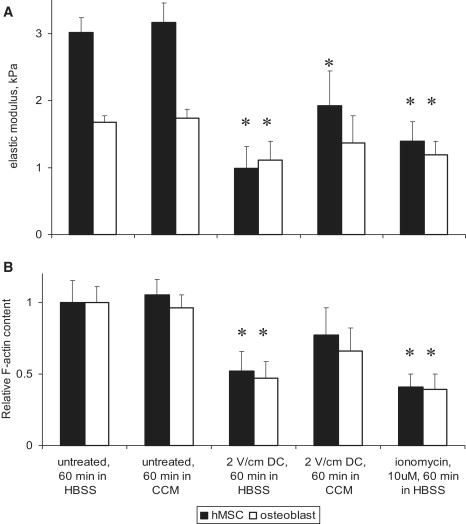
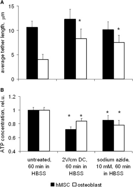
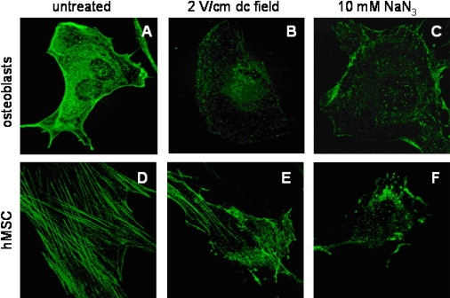
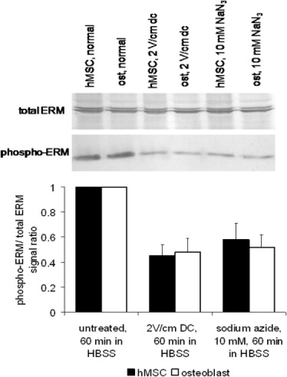
Cellular mechanics is known to play an important role in the cell homeostasis including proliferation, motility, and differentiation. Significant variation in the mechanical properties between different cell types suggests that control of the cell metabolism is feasible through manipulation of the cell mechanical parameters using external physical stimuli. We investigated the electrocoupling mechanisms of cellular biomechanics modulation by an electrical stimulation in two mechanically distinct cell types--human mesenchymal stem cells and osteoblasts. Application of a 2 V/cm direct current electric field resulted in approximately a twofold decrease in the cell elasticity and depleted intracellular ATP. Reduction in the ATP level led to inhibition of the linker proteins that are known to physically couple the cell membrane and cytoskeleton. The membrane separation from the cytoskeleton was confirmed by up to a twofold increase in the membrane tether length that was extracted from the cell membrane after an electrical stimulation. In comparison to human mesenchymal stem cells, the membrane-cytoskeleton attachment in osteoblasts was much stronger but, in response to the same electrical stimulation, the membrane detachment from the cytoskeleton was found to be more pronounced. The observed effects mediated by an electric field are cell type- and serum-dependent and can potentially be used for electrically assisted cell manipulation. An in-depth understanding and control of the mechanisms to regulate cell mechanics by external physical stimulus (e.g., electric field) may have great implications for stem cell-based tissue engineering and regenerative medicine.


Figures

Figure 1
Figure 1
Effect of electric field on cell cytoskeleton. Cells were exposed to a 2 V/cm dc field for 60 min in HBSS, and complete cell culture medium (CCM). (A) Cell cytoskeleton elasticity determined with AFM microindentation. The elastic modulus decreased significantly after an electrical stimulation or cell treatment with calcium ionophore in HBSS in both hMSCs and osteoblasts. Between 400–600 force-curves were acquired for each cell type and experimental condition. (B) Relative F-actin content in cells exposed to the electric field. It was obtained for each cell as a ratio of fluorescence signals from F-actin and the total protein, and then normalized to the corresponding control. The highest degree of actin depolymerization was observed in electrically stimulated cells in the absence of serum. Results represent the mean ± SE. Statistically different from respective controls (p < 0.05).
Figure 2
Figure 2
Actin cytoskeleton reorganization. Fluorescent images of osteoblasts are in the left column and hMSCs in the right column. Immunofluorescent images of a thin filamentous actin meshwork (red) and vinculins (green) showed that osteoblasts contain fewer and smaller focal adhesions (A). In contrast, hMSCs showed thick actin stress fibers, and multiple and large adhesion contacts (B). Actins were partially disassembled in both osteoblast (C) and hMSC (D) after cell exposure to a 2 V/cm field for 60 min in serum-free HBSS. Cells treatment with 10 μM ionomycin for 40 min in HBSS produced analogous actin dismantling effects both in osteoblast (E) and hMSC (F). When cells were placed in cell culture medium with serum, the electrical stimulation again caused redistribution of actins in osteoblast (G) and stem cells (H), but in a manner that was different than (C) and (D) without serum. Cell incubation in the complete culture medium for 60 min at 37°C after the dc field exposure resulted in a partial recovery of the actin structure in both osteoblasts (I) and hMSC (J). The arrows indicate the direction of the electric field applied.
Figure 3
Figure 3
Electrically induced changes in the plasma membrane mechanics. (A) Membrane tether length measured with laser optical tweezers. Much longer tethers could be extracted from the normal hMSC membrane than that of osteoblasts, likely due to a weaker membrane-cytoskeleton interaction in stem cells. The tether lengths increased in osteoblasts after cell exposure to a 2 V/cm field or 10 mM sodium azide and 10 mM 2-deoxyglucose. At least 30 tethers were measured for each cell type and experimental condition. (B) Intracellular ATP content determined with bioluminescent luciferin-luciferase assay. The ATP content was normalized to untreated samples. Results represent the mean ± SE. Statistically different from respective controls (p < 0.05).
Figure 4
Figure 4
ERM protein redistribution in response to an electric field. Osteoblasts are shown in the upper row, and hMSCs in the lower row. Immunofluorescently labeled ezrin, radixin, and moesin proteins were uniformly distributed across the osteoblast membrane, likely due to high density ERM binding sites on the dense actin meshwork contiguous to the membrane (A). In contrast, in normal hMSCs ERM linkers were found localized only along the stress fibers adjacent to the juxtaposed membrane (D). After cell stimulation with a 2 V/cm field for 60 min, the amount of active (phosphorylated membrane-bound) ERM proteins seemed to decrease in osteoblast (B) as well as in hMSC (E). Similar effect was achieved by ATP depletion using 10 mM sodium azide and 10 mM 2-deoxyglucose in osteoblasts (C) and hMSCs (F). All images are 100 × 100 μm in size.
Figure 5
Figure 5
Electrically induced ERM protein inhibition. Immunoblotting of total and phosphorylated ERM proteins in osteoblasts and hMSC. Phosphorylation of inactive ezrin, radixin, and moesin in the cell cytoplasm allows their binding to both actin and transmembrane proteins, and dephosphorylation inhibits the membrane-cytoskeleton linker function. After cell exposure to a 2 V/cm dc electric field in HBSS or metabolic inhibitors sodium azide and 2-deoxyglucose for 60 min, cell lysates were prepared, and equal amounts of proteins per well were separated by SDS-PAGE, and the total and phosphorylated ERM proteins were detected by Western blotting (top) and quantified by densitometry scanning. The data for phospho-ERM immunoblot signal relative to untreated cell control and then normalized to the total ERM signal is shown as mean ± SE from four independent experiments (bottom). All treatments were significantly different from their respective controls (p < 0.05).
Figure 6
Figure 6
Schematic for electrocoupling mechanisms of cell mechanics modulation by electric field. External electric field induces an increase in the cytosolic calcium concentration mediated either by Ca2+ influx through plasma membrane or Ca2+ release from intracellular store. An elevated intracellular Ca2+ level depolymerizes the F-actins and decrease the cell elasticity. If present (e.g., cell electrical exposure with serum), growth factors could bind to electrically redistributed plasma membrane receptors and trigger a local increase in actin polymerization. Redistribution of the membrane receptors and actins in response to an electric field may mediate serum-dependent cell electromigration. In addition, an electrical stimulation causes intracellular ATP depletion, for example, by ATP release, which in turn leads to inhibition of the ERM linkers' binding properties and their dissociation from the membrane and actin cytoskeleton. Resultant membrane separation from the cytoskeleton and effectively decreased membrane tension are attributed both to electrically induced downregulation of active ERM proteins and actin depolymerization. The exact details of these mechanisms may vary in different cell types.

Similar articles

Cited by

References

    1. Boscolo P., Di Gioacchino M., Di Giampaolo L., Antonucci A., Di Luzio S. Combined effects of electromagnetic fields on immune and nervous responses. Int. J. Immunopathol. Pharmacol. 2007;20:59–63. - PubMed
    1. Mycielska M.E., Djamgoz M.B. Cellular mechanisms of direct-current electric field effects: galvanotaxis and metastatic disease. J. Cell Sci. 2004;117:1631–1639. - PubMed
    1. Song B., Zhao M., Forrester J., McCaig C. Nerve regeneration and wound healing are stimulated and directed by an endogenous electrical field in vivo. J. Cell Sci. 2004;117:4681–4690. - PubMed
    1. Hotary K.B., Robinson K.R. Endogenous electrical currents and voltage gradients in Xenopus embryos and the consequences of their disruption. Dev. Biol. 1994;166:789–800. - PubMed
    1. Revest P.A., Jones H.C., Abbott N.J. Transendothelial electrical potential across pial vessels in anesthetized rats: a study of ion permeability and transport at the blood-brain barrier. Brain Res. 1994;652:76–82. - PubMed

Publication types

LinkOut - more resources