. 2009 Jan 23;136(2):249-60.
doi: 10.1016/j.cell.2008.11.017.

Induction of hepatitis by JNK-mediated expression of TNF-alpha

Affiliations

Induction of hepatitis by JNK-mediated expression of TNF-alpha

Madhumita Das et al. Cell. .

Abstract

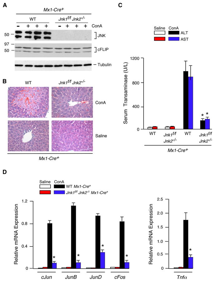
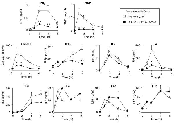
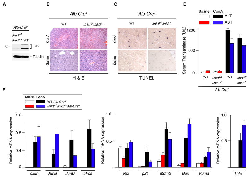
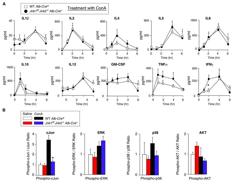
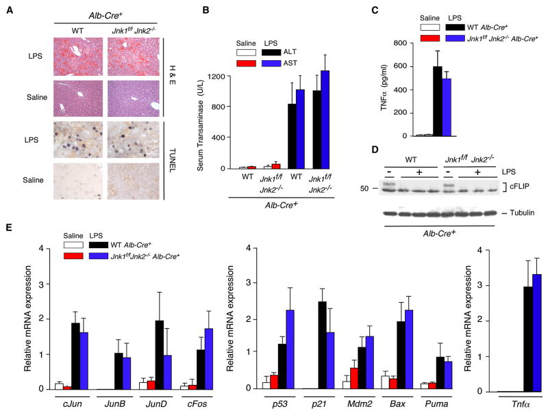
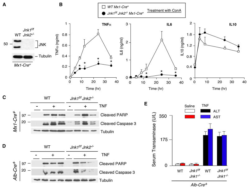
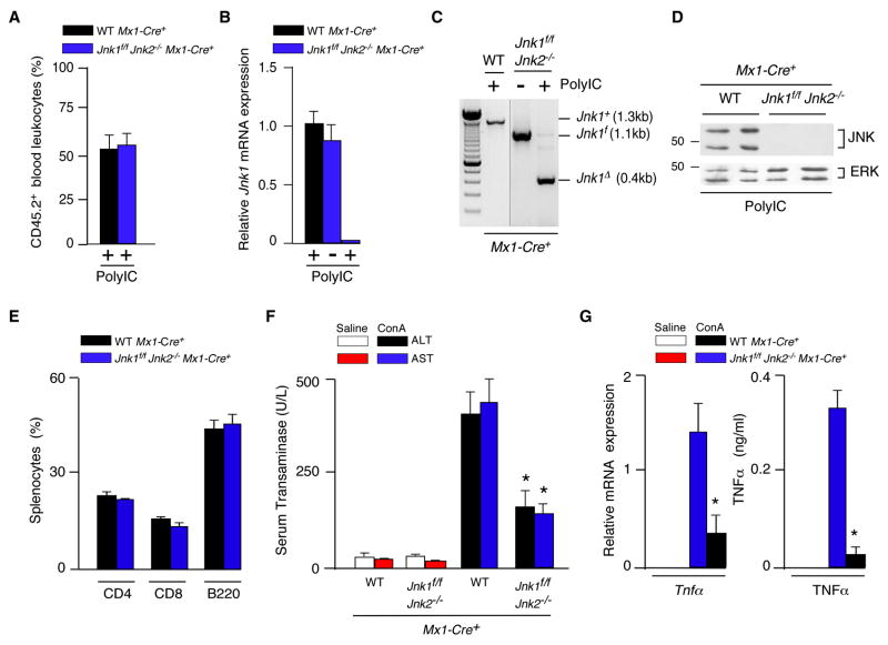
The c-Jun NH(2)-terminal kinase (JNK) signaling pathway has been implicated in the development of tumor necrosis factor (TNF)-dependent hepatitis. JNK may play a critical role in hepatocytes during TNF-stimulated cell death in vivo. To test this hypothesis, we examined the phenotype of mice with compound disruption of the Jnk1 and Jnk2 genes. Mice with loss of JNK1/2 expression in hepatocytes exhibited no defects in the development of hepatitis compared with control mice, whereas mice with loss of JNK1/2 in the hematopoietic compartment exhibited a profound defect in hepatitis that was associated with markedly reduced expression of TNF-alpha. These data indicate that JNK is required for TNF-alpha expression but not for TNF-alpha-stimulated death of hepatocytes. Indeed, TNF-alpha induced similar hepatic damage in both mice with hepatocyte-specific JNK1/2 deficiency and control mice. These observations confirm a role for JNK in the development of hepatitis but identify hematopoietic cells as the site of the essential function of JNK.


Figures

Figure 1
Figure 1. JNK-deficient mice are protected against ConA-induced hepatitis
(A) PolyIC-treated control mice (Mx1-Cre+) and JNK-deficient mice (Jnk1f/f Jnk2−/− Mx1-Cre+) were aged 4 weeks and then treated intravenously (8 hrs) with ConA or solvent (saline). Liver extracts were examined by immunoblot analysis using antibodes to JNK1/2, cFLIP, and α-Tubulin. (B) Representative H&E-stained liver sections prepared from control and JNK1/2-deficient mice treated (8 hrs) with ConA or solvent (saline) are presented. The amount of liver damage was quantitated (Figure S2C). (C) Serum transaminase activity in control and JNK1/2-deficient mice treated (8 hrs) with ConA or solvent (saline) was measured (mean ± SD; n = 6). Statistically significant differences between wild-type and JNK1/2-deficient mice are indicated (*, P < 0.01). (D) RNA was isolated from the liver of control and JNK1/2-deficient mice treated (8 hrs) with ConA or solvent (saline). The expression of mRNA was measured by quantitative RT-PCR assays. The mRNA expression was normalized to the amount of Gapdh mRNA and presented as the mean ± SD (n = 6). Statistically significant differences between wild-type and JNK1/2-deficient mice are indicated (*, P < 0.01).
Figure 2
Figure 2. JNK-deficient mice exhibit defects in the expression of serum cytokines
PolyIC-treated control mice (Mx1-Cre+) and JNK-deficient mice (Jnk1f/f Jnk2−/− Mx1-Cre+) were aged 4 weeks and then treated intravenously with ConA. The amount of serum cytokines post-treatment with ConA was measured by ELISA (mean ± SD; n = 7). Statistically significant differences between wild-type and JNK1/2-deficient mice are indicated (*, P < 0.05; **, P < 0.01).
Figure 3
Figure 3. JNK-deficiency in hepatocytes does not protect mice against ConA-induced hepatitis
(A) Liver extracts prepared from control mice (Alb-Cre+) and mice with hepatocyte-specific JNK-deficiency (Jnk1f/f Jnk2−/− Alb-Cre+) were examined by immunoblot analysis using antibodies to JNK1/2 and α-Tubulin. (B,C) Control mice (Alb-Cre+) and JNK-deficient mice (Jnk1f/f Jnk2−/− Alb-Cre+) were treated intravenously (8 hrs) with ConA or solvent (saline). Representative H&E-stained (B) and TUNEL- stained (C) liver sections prepared from control and JNK1/2-deficient mice are presented. (D) Serum transaminase activity in control and JNK-deficient mice after treatment (8 hrs) with ConA or solvent (saline) was measured (mean ± SD; n = 6). No statistically significant differences between wild-type and JNK1/2-deficient mice were detected. (E) RNA was isolated from the liver of control and JNK-deficient mice after treatment (8 hrs) with ConA or solvent (saline). The expression of mRNA was measured by quantitative RT-PCR assays (Taqman©). The mRNA expression was normalized to the amount of Gapdh mRNA and presented as the mean ± SD (n = 6). No statistically significant differences between wild-type and JNK1/2-deficient mice were detected.
Figure 4
Figure 4. Effect of hepatocyte-specific JNK-deficiency on serum cytokine expression
(A) Control mice (Alb-Cre+) and mice with hepatocyte-specific JNK-deficiency (Jnk1f/f Jnk2−/−Alb-Cre+) were treated intravenously with ConA. The amount of serum cytokines (IL1, IL2, IL4, IL5, IL6, IL10, IL12, GM-CSF, TNFα, and IFNγ) post-treatment with ConA was measured by ELISA (mean ± SD; n = 8). Statistically significant differences between wild-type and JNK1/2-deficient mice are indicated (*, P < 0.05). (B) The amount of total and phospho- cJun, ERK, p38 MAPK, and AKT in liver extracts at 8 hrs post-treatment of mice with ConA or saline was measured by multiplexed ELISA (mean ± SD; n = 5). Statistically significant differences between wild-type and JNK1/2-deficient mice are indicated (*, P < 0.01).
Figure 5
Figure 5. JNK-deficiency in hepatocytes does not protect mice against LPS-induced hepatitis
(A) Control mice (Alb-Cre+) and mice with hepatocyte-specific JNK-deficiency (Jnk1f/f Jnk2−/−Alb-Cre+) were treated intravenously (8 hrs) with LPS plus GalN or solvent (saline). Representative H&E- and TUNEL- stained liver sections prepared from control and JNK-deficient mice are presented. (B) Serum transaminase activity in control and JNK1/2-deficient mice after treatment (8 hrs) with LPS/GalN or solvent (saline) was measured (mean ± SD; n = 6). No statistically significant differences between wild-type and JNK1/2-deficient mice were detected. (C) The concentration of TNFα in the serum of control mice (Alb-Cre+) and JNK1/2-deficient mice (Jnk1f/f Jnk2−/− Alb-Cre+) was measured after treatment (1 hr) with LPS/GalN or solvent (saline) by ELISA (mean ± SD; n = 6). No statistically significant differences between wild-type and JNK1/2-deficient mice were detected. (D) Liver extracts prepared from control mice (Alb-Cre+) and JNK1/2-deficient mice (Jnk1f/f Jnk2−/− Alb-Cre+) were examined by immunoblot analysis using antibodies to cFLIP and α-Tubulin. (E) RNA was isolated from the liver of control and JNK-deficient mice after treatment (8 hrs) with LPS/GalN or solvent (saline). The expression of mRNA was measured by quantitative RT-PCR assays. The mRNA expression was normalized to the amount of Gapdh mRNA and presented as the mean ± SD (n = 6). No statistically significant differences between wild-type and JNK1/2-deficient mice were detected.
Figure 6
Figure 6. JNK-deficiency inhibits TNFα expression, but not TNFα-induced liver damage
(A,B) Bone marrow-derived macrophages prepared from polyIC-treated control mice (Mx1-Cre+) and JNK-deficient mice (Jnk1f/f Jnk2−/− Mx1-Cre+) were examined by immunoblot analysis using antibodies to JNK1/2 and α-Tubulin. The macrophages were treated with ConA and cytokines in the culture medium were measured by ELISA (mean ± SD; n = 7). Statistically significant differences between wild-type and JNK1/2-deficient mice are indicated (*, P < 0.01). (C,D) PolyIC-treated control mice (Mx1-Cre+) and JNK-deficient mice (Jnk1f/f Jnk2−/− Mx1-Cre+) (C) or control mice (Alb-Cre+) and mice with hepatocyte-specific JNK-deficiency (Jnk1f/f Jnk2−/−Alb-Cre+) (D) were treated intravenously (8 hrs) with TNFα plus GalN or solvent (saline). Liver extracts were examined by immunoblot analysis using antibodies to capsase-cleaved PARP, cleaved caspase-3, and α-Tubulin. (E) Serum transaminase activity in control and JNK1/2-deficient mice after treatment (8 hrs) with LPS/GalN or solvent (saline) was measured (mean ± SD; n = 6). No statistically significant differences between wild-type and JNK1/2-deficient mice were detected.
Figure 7
Figure 7. JNK-deficiency in hematopoietic cells protects mice against hepatitis
(A) Competitive bone marrow transplantation assays were performed using lethally-irradiated wild-type B6.SJL mice transplanted with an equal number of bone marrow cells isolated from polyIC-treated (4 weeks) control B6.SJL (CD45.1) mice plus Jnk1f/f Jnk2−/− Mx1-Cre+ or wild-type Mx1-Cre+ C57BL/6J (CD45.2) mice. Peripheral blood leukocytes were stained with antibodies to CD45.1 and CD45.2 at 6 months post-transplantation by flow cytometry (mean ± SD; n = 3). (B–E) Lethally-irradiated wild-type mice were transplanted with bone marrow from polyIC-treated control mice (Mx1-Cre+) or JNK1/2-deficient mice (Jnk1f/f Jnk2−/− Mx1-Cre+) mice. Peripheral blood leukocytes were isolated and Jnk1 mRNA was examined by quantitative Taqman© RT-PCR analysis (B) and genomic DNA was genotyped by PCR analysis using amplimers to detect the Jnk1+, Jnk1f, and Jnk1Δ alleles (C). Splenocytes were examined by immunoblot analysis using antibodies to JNK1/2 and ERK1/2 (D) and sub-populations of splenocytes were examined by flow cytomtetry (E). (F) Mice at 6 months post-transplantation were treated intravenously (8 hrs) with ConA or solvent (saline) and serum transaminase activity (ALT and AST) was measured (mean ± SD; n = 6). Statistically significant differences between mice transplanted with control and JNK1/2-deficient bone marrow are indicated (*, P < 0.01). (G) Mice at 6 months post-transplantation were treated intravenously with ConA or solvent (saline). The expression of Gapdh, and Tnfα mRNA in the liver was measured by quantitative RT-PCR assays (Taqman©) at 8 hr. post-injection (left panel). The mRNA expression in each sample was normalized to the amount of Gapdh mRNA and presented as the mean ± SD (n = 6). The concentration of TNFα in the blood was measured at 1 hr. post-injection by ELISA and is presented as the mean ± SD (n = 7) (right panel). Statistically significant differences between mice transplanted with control and JNK1/2-deficient bone marrow are indicated (*, P < 0.01).

References

    1. Adhikari A, Xu M, Chen ZJ. Ubiquitin-mediated activation of TAK1 and IKK. Oncogene. 2007;26:3214–3226. - PubMed
    1. Alves-Guerra MC, Rousset S, Pecqueur C, Mallat Z, Blanc J, Tedgui A, Bouillaud F, Cassard-Doulcier AM, Ricquier D, Miroux B. Bone marrow transplantation reveals the in vivo expression of the mitochondrial uncoupling protein 2 in immune and nonimmune cells during inflammation. J Biol Chem. 2003;278:42307–42312. - PubMed
    1. Bonder CS, Ajuebor MN, Zbytnuik LD, Kubes P, Swain MG. Essential role for neutrophil recruitment to the liver in concanavalin A-induced hepatitis. J Immunol. 2004;172:45–53. - PubMed
    1. Chan FK, Lenardo MJ. A crucial role for p80 TNF-R2 in amplifying p60 TNF-R1 apoptosis signals in T lymphocytes. Eur J Immunol. 2000;30:652–660. - PubMed
    1. Chang L, Kamata H, Solinas G, Luo JL, Maeda S, Venuprasad K, Liu YC, Karin M. The E3 ubiquitin ligase itch couples JNK activation to TNFalpha-induced cell death by inducing c-FLIP(L) turnover. Cell. 2006;124:601–613. - PubMed

Publication types

MeSH terms

Substances