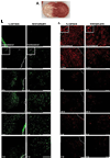
TTC, fluoro-Jade B and NeuN staining confirm evolving phases of infarction induced by middle cerebral artery occlusion
- PMID: 19167427
- PMCID: PMC2674851
- DOI: 10.1016/j.jneumeth.2008.12.028
TTC, fluoro-Jade B and NeuN staining confirm evolving phases of infarction induced by middle cerebral artery occlusion
Abstract
Considerable debate exists in the literature on how best to measure infarct damage and at what point after middle cerebral artery occlusion (MCAO) infarct is histologically complete. As many researchers are focusing on more chronic endpoints in neuroprotection studies it is important to evaluate histological damage at later time points to ensure that standard methods of tissue injury measurement are accurate. To compare tissue viability at both acute and sub-acute time points, we used 2,3,5-triphenyltetrazolium chloride (TTC), Fluoro-Jade B, and NeuN staining to examine the evolving phases of infarction induced by a 90-min MCAO in mice. Stroke outcomes were examined at 1.5h, 6h, 12h, 24h, 3d, and 7d after MCAO. There was a time-dependent increase in infarct volume from 1.5h to 24h in the cortex, followed by a plateau from 24h to 7d after stroke. Striatal infarcts were complete by 12h. Fluoro-Jade B staining peaked at 24h and was minimal by 7d. Our results indicated that histological damage as measured by TTC and Fluoro-Jade B reaches its peak by 24h after stroke in a reperfusion model of MCAO in mice. TTC staining can be accurately performed as late as 7d after stroke. Neurological deficits do not correlate with the structural lesion but rather transient impairment of function. As the infarct is complete by 24h and even earlier in the striatum, even the most efficacious neuroprotective therapies are unlikely to show any efficacy if given after this point.
Figures


Similar articles
-
Temporary focal ischemia in the mouse: technical aspects and patterns of Fluoro-Jade evident neurodegeneration.Brain Res. 2005 Apr 25;1042(1):29-36. doi: 10.1016/j.brainres.2005.02.021. Brain Res. 2005. PMID: 15823250
-
Mouse model of intraluminal MCAO: cerebral infarct evaluation by cresyl violet staining.J Vis Exp. 2012 Nov 6;(69):4038. doi: 10.3791/4038. J Vis Exp. 2012. PMID: 23168377 Free PMC article.
-
Sigma receptor activation reduces infarct size at 24 hours after permanent middle cerebral artery occlusion in rats.Curr Neurovasc Res. 2006 May;3(2):89-98. doi: 10.2174/156720206776875849. Curr Neurovasc Res. 2006. PMID: 16719792
-
Left-right asymmetry influenced the infarct volume and neurological dysfunction following focal middle cerebral artery occlusion in rats.Brain Behav. 2018 Dec;8(12):e01166. doi: 10.1002/brb3.1166. Epub 2018 Nov 19. Brain Behav. 2018. PMID: 30451395 Free PMC article.
-
Infiltration of invariant natural killer T cells occur and accelerate brain infarction in permanent ischemic stroke in mice.Neurosci Lett. 2016 Oct 28;633:62-68. doi: 10.1016/j.neulet.2016.09.010. Epub 2016 Sep 13. Neurosci Lett. 2016. PMID: 27637387
Cited by
-
Sulfide catabolism ameliorates hypoxic brain injury.Nat Commun. 2021 May 25;12(1):3108. doi: 10.1038/s41467-021-23363-x. Nat Commun. 2021. PMID: 34035265 Free PMC article.
-
Inhibition of the group I mGluRs reduces acute brain damage and improves long-term histological outcomes after photothrombosis-induced ischaemia.ASN Neuro. 2013 Jul 11;5(3):195-207. doi: 10.1042/AN20130002. ASN Neuro. 2013. PMID: 23772679 Free PMC article.
-
Dodecafluoropentane Emulsion (DDFPe) Decreases Stroke Size and Improves Neurological Scores in a Permanent Occlusion Rat Stroke Model.Open Neurol J. 2014 Dec 30;8:27-33. doi: 10.2174/1874205X01408010027. eCollection 2014. Open Neurol J. 2014. PMID: 25674164 Free PMC article.
-
Neuroprotective Effects of Musk of Muskrat on Transient Focal Cerebral Ischemia in Rats.Evid Based Complement Alternat Med. 2019 Jun 25;2019:9817949. doi: 10.1155/2019/9817949. eCollection 2019. Evid Based Complement Alternat Med. 2019. PMID: 31341507 Free PMC article.
-
KLF4 alleviates cerebral vascular injury by ameliorating vascular endothelial inflammation and regulating tight junction protein expression following ischemic stroke.J Neuroinflammation. 2020 Apr 7;17(1):107. doi: 10.1186/s12974-020-01780-x. J Neuroinflammation. 2020. PMID: 32264912 Free PMC article.
References
-
- Adkins DL, Voorhies AC, Jones TA. Behavioral and neuroplastic effects of focal endothelin-1 induced sensorimotor cortex lesions. Neuroscience. 2004;128:473–486. - PubMed
-
- Ajmo CT, Jr, Vernon DO, Collier L, Pennypacker KR, Cuevas J. Sigma receptor activation reduces infarct size at 24 hours after permanent middle cerebral artery occlusion in rats. Curr Neurovasc Res. 2006;3:89–98. - PubMed
-
- Alexis NE, Back T, Zhao W, Dietrich WD, Watson BD, Ginsberg MD. Neurobehavioral consequences of induced spreading depression following photothrombotic middle cerebral artery occlusion. Brain Res. 1996;706:273–282. - PubMed
-
- Aspey BS, Cohen S, Patel Y, Terruli M, Harrison MJ. Middle cerebral artery occlusion in the rat: consistent protocol for a model of stroke. Neuropathol Appl Neurobiol. 1998;24:487–497. - PubMed
-
- Baird AE, Benfield A, Schlaug G, Siewert B, Lovblad KO, Edelman RR, Warach S. Enlargement of human cerebral ischemic lesion volumes measured by diffusion-weighted magnetic resonance imaging. Ann Neurol. 1997;41:581–589. - PubMed
Publication types
MeSH terms
Substances
Grants and funding
LinkOut - more resources
Full Text Sources
Research Materials

