

Persistent working memory dysfunction following traumatic brain injury: evidence for a time-dependent mechanism
- PMID: 19167462
- PMCID: PMC4264540
- DOI: 10.1016/j.neuroscience.2008.12.050
Persistent working memory dysfunction following traumatic brain injury: evidence for a time-dependent mechanism
Abstract
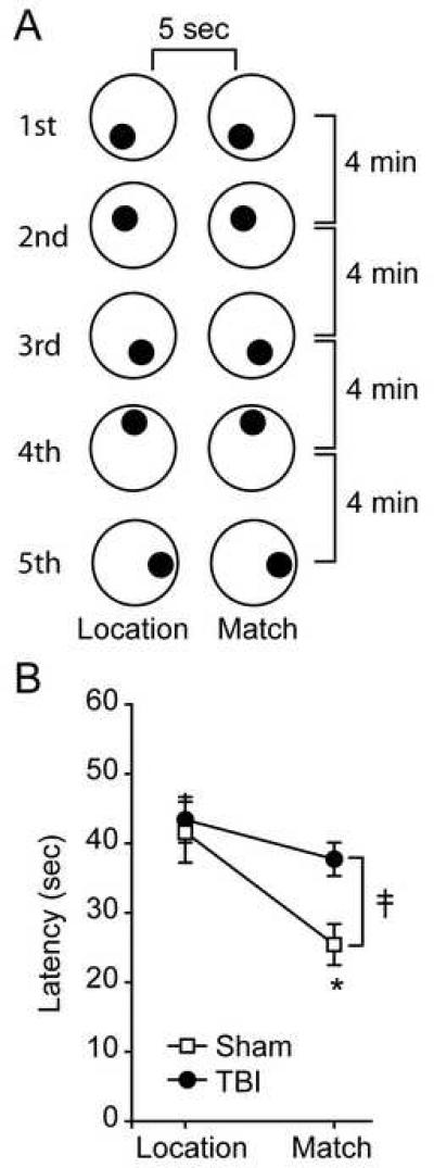
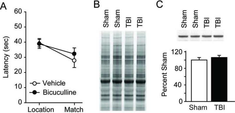
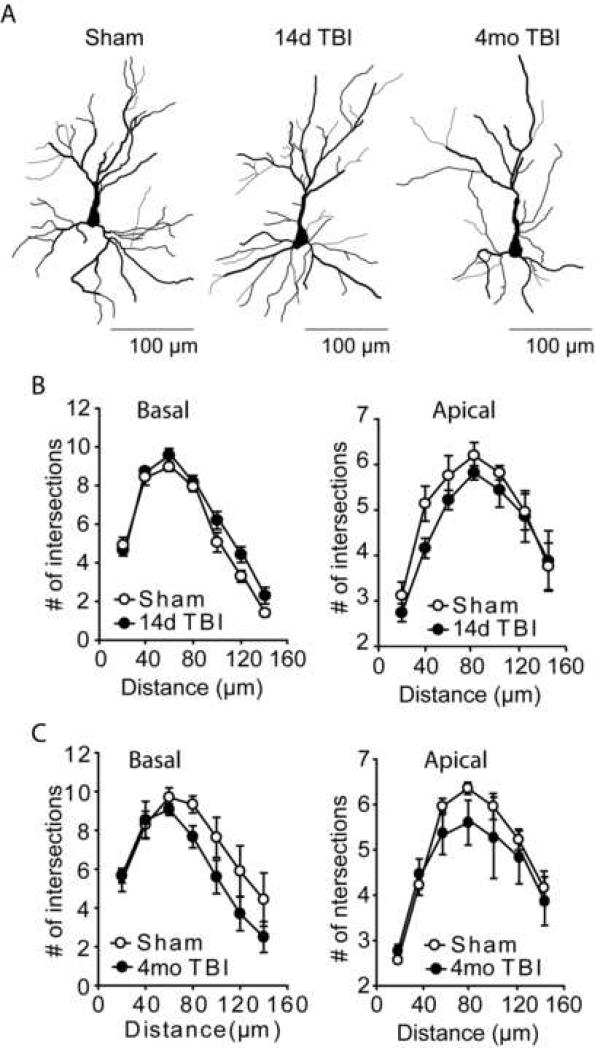
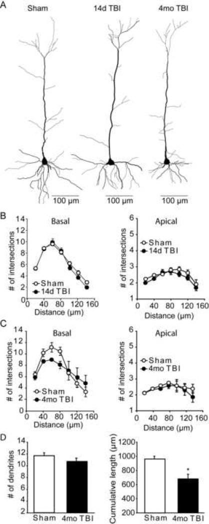
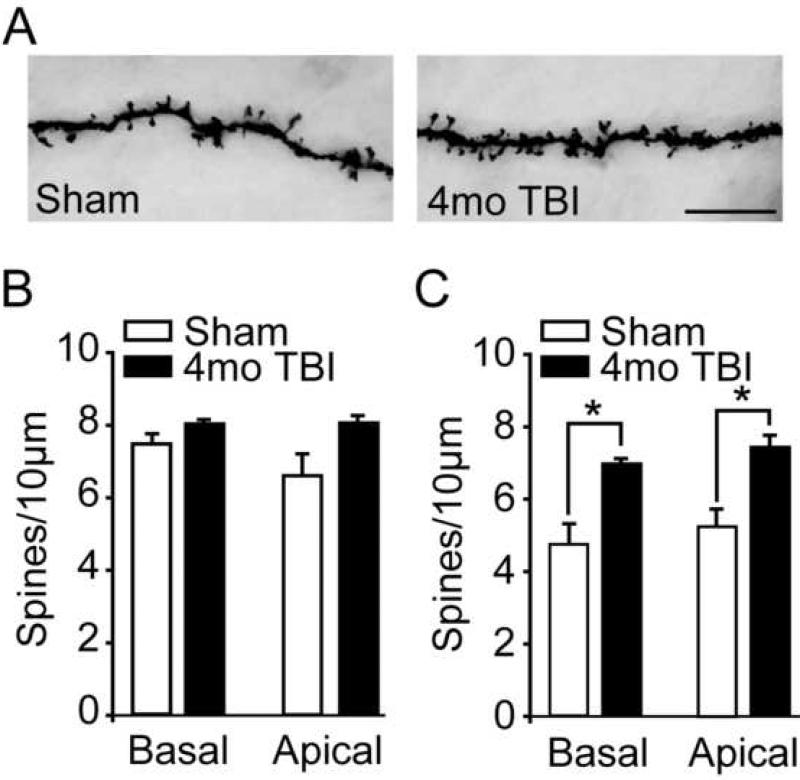
The prefrontal cortex is highly vulnerable to traumatic brain injury (TBI) resulting in the dysfunction of many high-level cognitive and executive functions such as planning, information processing speed, language, memory, attention, and perception. All of these processes require some degree of working memory. Interestingly, in many cases, post-injury working memory deficits can arise in the absence of overt damage to the prefrontal cortex. Recently, excess GABA-mediated inhibition of prefrontal neuronal activity has been identified as a contributor to working memory dysfunction within the first month following cortical impact injury of rats. However, it has not been examined if these working memory deficits persist, and if so, whether they remain amenable to treatment by GABA antagonism. Our findings show that working memory dysfunction, assessed using both the delay match-to-place and delayed alternation T-maze tasks, following lateral cortical impact injury persists for at least 16 weeks post-injury. These deficits were found to be no longer the direct result of excess GABA-mediated inhibition of medial prefrontal cortex neuronal activity. Golgi staining of prelimbic pyramidal neurons revealed that TBI causes a significant shortening of layers V/VI basal dendrite arbors by 4 months post-injury, as well as an increase in the density of both basal and apical spines in these neurons. These changes were not observed in animals 14 days post-injury, a time point at which administration of GABA receptor antagonists improves working memory function. Taken together, the present findings, along with previously published reports, suggest that temporal considerations must be taken into account when designing mechanism-based therapies to improve working memory function in TBI patients.
Figures

Similar articles
-
Altered adrenergic receptor signaling following traumatic brain injury contributes to working memory dysfunction.Neuroscience. 2011 Jan 13;172:293-302. doi: 10.1016/j.neuroscience.2010.10.048. Epub 2010 Oct 23. Neuroscience. 2011. PMID: 20974230 Free PMC article.
-
Effects of selective estrogen receptor modulators on allocentric working memory performance and on dendritic spines in medial prefrontal cortex pyramidal neurons of ovariectomized rats.Horm Behav. 2012 Apr;61(4):512-7. doi: 10.1016/j.yhbeh.2012.01.010. Epub 2012 Jan 21. Horm Behav. 2012. PMID: 22285935
-
Prefrontal cortical GABA modulation of spatial reference and working memory.Int J Neuropsychopharmacol. 2014 Oct 31;18(2):pyu013. doi: 10.1093/ijnp/pyu013. Int J Neuropsychopharmacol. 2014. PMID: 25552433 Free PMC article.
-
Selective alterations in prefrontal cortical GABA neurotransmission in schizophrenia: a novel target for the treatment of working memory dysfunction.Psychopharmacology (Berl). 2004 Jun;174(1):143-50. doi: 10.1007/s00213-003-1673-x. Epub 2003 Dec 9. Psychopharmacology (Berl). 2004. PMID: 15205885 Review.
-
Beyond working memory: the role of persistent activity in decision making.Trends Cogn Sci. 2010 May;14(5):216-22. doi: 10.1016/j.tics.2010.03.006. Epub 2010 Apr 8. Trends Cogn Sci. 2010. PMID: 20381406 Free PMC article. Review.
Cited by
-
Early Life Stress Exacerbates Outcome after Traumatic Brain Injury.J Neurotrauma. 2021 Mar;38(5):555-565. doi: 10.1089/neu.2020.7267. Epub 2020 Sep 16. J Neurotrauma. 2021. PMID: 32862765 Free PMC article.
-
Progressive long-term spatial memory loss following repeat concussive and subconcussive brain injury in mice, associated with dorsal hippocampal neuron loss, microglial phenotype shift, and vascular abnormalities.Eur J Neurosci. 2021 Sep;54(5):5844-5879. doi: 10.1111/ejn.14711. Epub 2020 Mar 12. Eur J Neurosci. 2021. PMID: 32090401 Free PMC article.
-
Involvement of the glycogen synthase kinase-3 signaling pathway in TBI pathology and neurocognitive outcome.PLoS One. 2011;6(9):e24648. doi: 10.1371/journal.pone.0024648. Epub 2011 Sep 15. PLoS One. 2011. PMID: 21935433 Free PMC article.
-
Pathophysiology and Treatment of Memory Dysfunction After Traumatic Brain Injury.Curr Neurol Neurosci Rep. 2017 Jul;17(7):52. doi: 10.1007/s11910-017-0762-x. Curr Neurol Neurosci Rep. 2017. PMID: 28500417 Free PMC article. Review.
-
Chronic Cognitive Dysfunction after Traumatic Brain Injury Is Improved with a Phosphodiesterase 4B Inhibitor.J Neurosci. 2016 Jul 6;36(27):7095-108. doi: 10.1523/JNEUROSCI.3212-15.2016. J Neurosci. 2016. PMID: 27383587 Free PMC article.
References
-
- Amat J, Baratta MV, Paul E, Bland ST, Watkins LR, Maier SF. (Medial prefrontal cortex determines how stressor controllability affects behavior and dorsal raphe nucleus. Nat Neurosci. 2005;8:365–371. - PubMed
-
- Arnsten AF. (Catecholamine regulation of the prefrontal cortex. J Psychopharmacol. 1997;11:151–162. - PubMed
-
- Baddeley A. (Working memory. Science. 1992;255:556–559. - PubMed
-
- Blum S, Hebert AE, Dash PK. (A role for the prefrontal cortex in recall of recent and remote memories. Neuroreport. 2006;17:341–344. - PubMed
-
- Bramlett HM, Dietrich WD. (Progressive damage after brain and spinal cord injury: pathomechanisms and treatment strategies. Prog Brain Res. 2007;161:125–141. - PubMed
Publication types
MeSH terms
Substances
Grants and funding
LinkOut - more resources
Full Text Sources
Other Literature Sources
Medical
