

Potential role of chemerin in recruitment of plasmacytoid dendritic cells to diseased skin
- PMID: 19168032
- PMCID: PMC6289204
- DOI: 10.1016/j.bbrc.2009.01.071
Potential role of chemerin in recruitment of plasmacytoid dendritic cells to diseased skin
Abstract
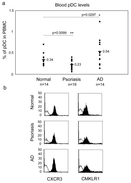
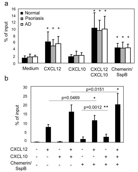
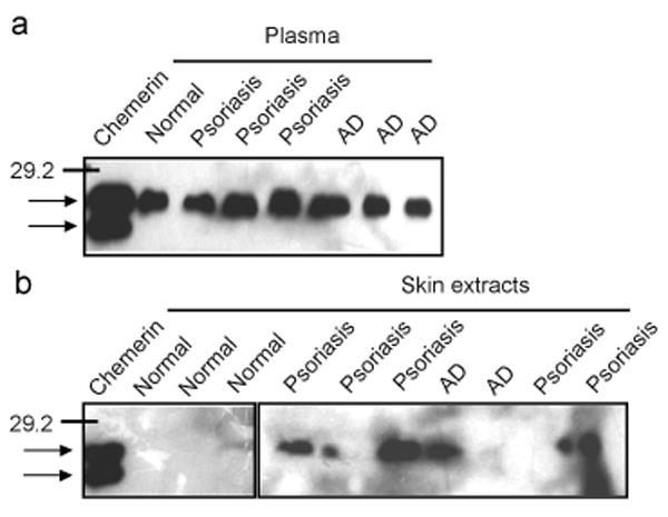
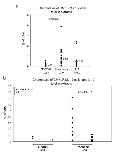
Interferon alpha-producing plasmacytoid dendritic cells (pDC) are crucial contributors to pro-inflammatory or tolerogenic immune responses and are important in autoimmune diseases such as psoriasis. pDC accumulate in the lesional skin of psoriasis patients, but are rarely found in the affected skin of patients with atopic dermatitis (AD). While homeostatic chemokine CXCL12 and inducible pro-inflammatory CXCR3 chemokine ligands may regulate pDC influx to psoriatic skin, the mechanism responsible for selective pDC recruitment in psoriasis vs. AD remains unknown. Circulating pDC from normal donors express a limited number of chemoattractant receptors, including CXCR3 and CMKLR1 (chemokine-like receptor 1). In this work, we demonstrate that circulating pDC from normal donors as well as psoriasis and AD patients express similar levels of CXCR3 and responded similarly in functional migration assays to CXCL10. We next found that blood pDC from normal, AD, and psoriasis patients express functional CMKLR1. In contrast to normal skin, however, lesional skin from psoriasis patients contains the active form of the CMKLR1 ligand chemerin. Furthermore, in affected skin from psoriatic patients the level of active chemerin was generally higher than in AD skin. Taken together, these results indicate that local generation of active chemerin may contribute to pDC recruitment to psoriatic skin.
Figures
Similar articles
-
Chemerin and the recruitment of NK cells to diseased skin.Acta Biochim Pol. 2009;56(2):355-60. Epub 2009 Jun 18. Acta Biochim Pol. 2009. PMID: 19543554 Free PMC article.
-
Chemerin expression marks early psoriatic skin lesions and correlates with plasmacytoid dendritic cell recruitment.J Exp Med. 2009 Jan 16;206(1):249-58. doi: 10.1084/jem.20080129. Epub 2008 Dec 29. J Exp Med. 2009. PMID: 19114666 Free PMC article.
-
The histamine H4 receptor is highly expressed on plasmacytoid dendritic cells in psoriasis and histamine regulates their cytokine production and migration.J Invest Dermatol. 2011 Aug;131(8):1668-76. doi: 10.1038/jid.2011.72. Epub 2011 May 26. J Invest Dermatol. 2011. PMID: 21614010
-
Immune functions and recruitment of plasmacytoid dendritic cells in psoriasis.Autoimmunity. 2010 Apr;43(3):215-9. doi: 10.3109/08916930903510906. Autoimmunity. 2010. PMID: 20166874 Review.
-
The role of dendritic cell subtypes in the pathophysiology of atopic dermatitis.J Am Acad Dermatol. 2005 Aug;53(2 Suppl 2):S171-6. doi: 10.1016/j.jaad.2005.04.060. J Am Acad Dermatol. 2005. PMID: 16021172 Review.
Cited by
-
Expression of human chemerin induces insulin resistance in the skeletal muscle but does not affect weight, lipid levels, and atherosclerosis in LDL receptor knockout mice on high-fat diet.Diabetes. 2010 Nov;59(11):2898-903. doi: 10.2337/db10-0362. Epub 2010 Aug 19. Diabetes. 2010. PMID: 20724582 Free PMC article.
-
Regulatory dendritic cells: there is more than just immune activation.Front Immunol. 2012 Sep 4;3:274. doi: 10.3389/fimmu.2012.00274. eCollection 2012. Front Immunol. 2012. PMID: 22969767 Free PMC article.
-
Staphylococcus aureus Proteases: Orchestrators of Skin Inflammation.DNA Cell Biol. 2024 Oct;43(10):483-491. doi: 10.1089/dna.2024.0134. Epub 2024 Jul 3. DNA Cell Biol. 2024. PMID: 38957987 Review.
-
Regulation of chemerin chemoattractant and antibacterial activity by human cysteine cathepsins.J Immunol. 2011 Aug 1;187(3):1403-10. doi: 10.4049/jimmunol.1002352. Epub 2011 Jun 29. J Immunol. 2011. PMID: 21715684 Free PMC article.
-
Altered dendritic cell functions in autoimmune diseases: distinct and overlapping profiles.Nat Rev Rheumatol. 2016 Dec;12(12):703-715. doi: 10.1038/nrrheum.2016.147. Epub 2016 Sep 22. Nat Rev Rheumatol. 2016. PMID: 27652503 Review.
References
-
- Liu YJ. IPC: professional type 1 interferon-producing cells and plasmacytoid dendritic cell precursors. Annu Rev Immunol. 2005;23:275–306. - PubMed
-
- Yoneyama H, Matsuno K, Zhang Y, Nishiwaki T, Kitabatake M, Ueha S, Narumi S, Morikawa S, Ezaki T, Lu B, Gerard C, Ishikawa S, Matsushima K. Evidence for recruitment of plasmacytoid dendritic cell precursors to inflamed lymph nodes through high endothelial venules. Int Immunol. 2004;16:915–928. - PubMed
-
- Penna G, Sozzani S, Adorini L. Cutting edge: selective usage of chemokine receptors by plasmacytoid dendritic cells. J Immunol. 2001;167:1862–1866. - PubMed
-
- Vanbervliet B, Bendriss-Vermare N, Massacrier C, Homey B, de Bouteiller O, Briere F, Trinchieri G, Caux C. The inducible CXCR3 ligands control plasmacytoid dendritic cell responsiveness to the constitutive chemokine stromal cell-derived factor 1 (SDF-1)/CXCL12. J Exp Med. 2003;198:823–830. - PMC - PubMed
-
- Zabel BA, Silverio AM, Butcher EC. Chemokine-like receptor 1 expression and chemerin-directed chemotaxis distinguish plasmacytoid from myeloid dendritic cells in human blood. J Immunol. 2005;174:244–251. - PubMed
Publication types
MeSH terms
Substances
Grants and funding
LinkOut - more resources
Full Text Sources
Medical
Molecular Biology Databases
Miscellaneous
