. 2009 Feb 17;106(7):2353-8.
doi: 10.1073/pnas.0812801106. Epub 2009 Jan 23.

Anthracycline chemotherapy inhibits HIF-1 transcriptional activity and tumor-induced mobilization of circulating angiogenic cells

Affiliations

Anthracycline chemotherapy inhibits HIF-1 transcriptional activity and tumor-induced mobilization of circulating angiogenic cells

KangAe Lee et al. Proc Natl Acad Sci U S A. .

Retraction in

Abstract

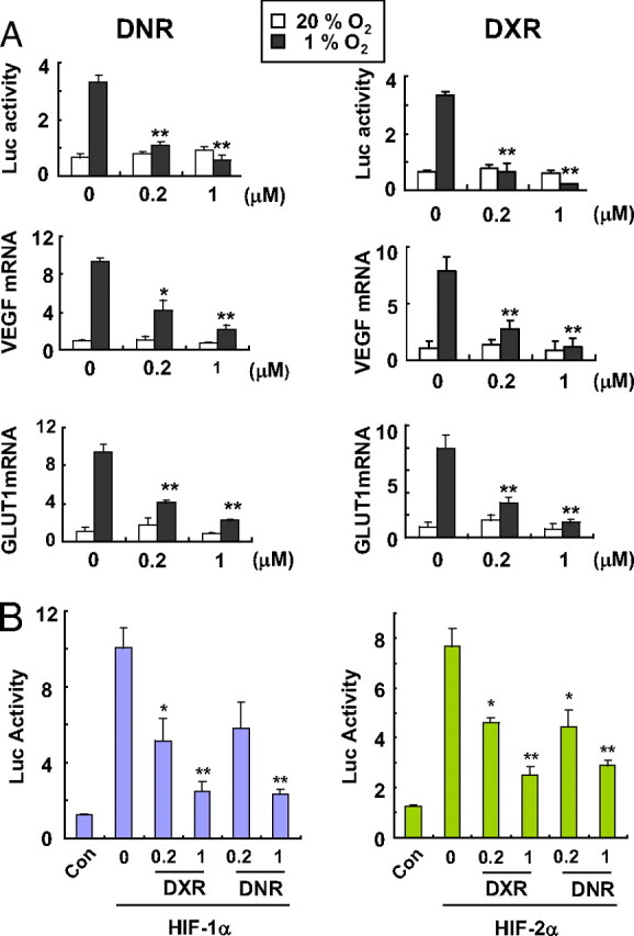
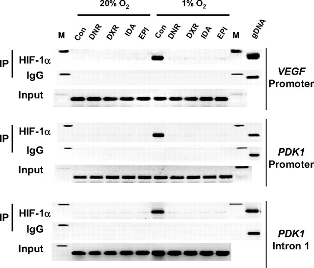
Using a cell-based reporter gene assay, we screened a library of drugs in clinical use and identified the anthracycline chemotherapeutic agents doxorubicin and daunorubicin as potent inhibitors of hypoxia-inducible factor 1 (HIF-1)-mediated gene transcription. These drugs inhibited HIF-1 by blocking its binding to DNA. Daily administration of doxorubicin or daunorubicin potently inhibited the transcription of a HIF-1-dependent reporter gene as well as endogenous HIF-1 target genes encoding vascular endothelial growth factor, stromal-derived factor 1, and stem cell factor in tumor xenografts. CXCR4(+)/sca1(+), VEGFR2(+)/CD34(+), and VEGFR2(+)/CD117(+) bone-marrow derived cells were increased in the peripheral blood of SCID mice bearing prostate cancer xenografts but not in tumor-bearing mice treated for 5 days with doxorubicin or daunorubicin, which dramatically reduced tumor vascularization. These results provide a molecular basis for the antiangiogenic effect of anthracycline therapy and have important implications for refining the use of these drugs to treat human cancer more effectively.


Conflict of interest statement

The authors declare no conflict of interest.

Figures

Fig. 1.
Fig. 1.
Inhibition of HIF-1 transcriptional activity by anthracyclines. (A) effect of DNR or DXR on hypoxia-induced HIF-1 activity. (Top) HIF-1-dependent Luc activity was determined in Hep3B-c1 cells treated with 0, 0.2, or 1 μM DNR or DXR under 20% (open bars) or 1% (filled bars) O2 for 24 h. Cells were lysed and analyzed for the ratio of firefly to Renilla Luc activity. (Middle and Bottom) HEK293 cells were exposed to 20% (open bars) or 1% (filled bars) O2 for 24 h in the presence of 0, 0.2, or 1 μM DNR or DXR. Total RNA was isolated for determination of VEGF (Middle) and GLUT1 (Bottom) mRNA levels by qRT-PCR. The mRNA levels were normalized to the levels of 18S rRNA in each sample, and each value was expressed relative to the levels in vehicle-treated cells exposed to 20% O2. Mean ± SEM are plotted (n = 4). *, P < 0.05; **, P < 0.01 (Student's t test). (B) effect of DNR or DXR on Luc activity mediated by cotransfection of expression vector encoding HIF-1α (Left, purple bars) or HIF-2α (Right, green bars). Mean ± SEM are plotted (n = 3). *, P < 0.05; **, P < 0.01 (Student's t test).
Fig. 2.
Fig. 2.
Analysis of HIF-1 DNA-binding activity by chromatin immunoprecipitation (IP) assay. HEK293 cells were exposed to 1% or 20% O2 in the presence of vehicle control (Con) or 1 μM DNR, DXR, EPI, or IDA for 20 h. Input DNA was isolated from an aliquot of lysate before IP, and lysates were then divided between anti-HIF-1α antibodies and rabbit IgG for IP. PCR was performed using the immunoprecipitates as template to amplify VEGF promoter, PDK1 promoter, and PDK1 intron 1 sequences. PCR products were analyzed by 2% agarose gel electrophoresis and ethidium bromide staining. M, 200-bp size marker.
Fig. 3.
Fig. 3.
Effect of DNR or DXR treatment on HIF-1 transcriptional activity in Hep3B-c1 tumor xenografts. (A) Mice bearing 200-mm3 Hep3B-c1 tumor xenografts were administered vehicle (blue), DNR (1 mg/kg per day, red), or DXR (1 mg/kg per day, green) by tail vein injection (red arrows). Tumor volume and body weight were monitored twice weekly. (B) HRE-driven Luc activity was determined in xenografts by Xenogen imaging before treatment (day 27, Upper) and 4 h after treatment on day 30, Lower). Mice were killed 4 h after treatment on day 31, and tumors from vehicle-, DNR-, and DXR-treated mice were collected to analyze HIF-1α protein levels (C) and VEGF, GLUT1, HK1, and HK2 mRNA levels (D), with mean ± SEM (n = 3) shown. *, P < 0.05; **, P < 0.01 (Student's t test).
Fig. 4.
Fig. 4.
Effect of DNR or DXR on tumor growth and mobilization of CACs into peripheral blood of mice bearing PC-3 prostate cancer xenografts. (A) PC-3 tumors were grown to a volume of ≈100 mm3, and mice were treated daily with vehicle (black), DNR (0.5 mg/kg, red; 1.5 mg/kg, blue), or DXR (0.5 mg/kg, green; 1.5 mg/kg, yellow) for 5 days (red arrows). Tumor volume was monitored twice weekly. (B) CACs were determined by using flow cytometry. Peripheral blood samples were collected 4 h after the last treatment, and the percentage of cells that were CXCR4+/sca1+, VEGFR2+/CD34+, or VEGFR2+/CD117+ was determined (mean ± SEM shown). (C) VEGF, SDF1, and SCF mRNA levels were determined in tumors from mice treated with vehicle (black), DNR (0.5 mg/kg, red; 1.5 mg/kg, blue), or DXR (0.5 mg/kg, green; 1.5 mg/kg, yellow). Mean ± SEM are shown (n = 4). *, P < 0.05; **, P < 0.01 (Student's t test). (D) Mice with (+) or without (−) PC-3 tumors of ≈100 mm3 were treated with vehicle (−) or 0.5 mg/kg DXR (+) for 5 days. Peripheral blood was collected 4 h after the last dose, and serum samples were analyzed for SDF-1α protein levels by ELISA; mean ± SEM are shown (n = 4). *, P < 0.01 vs. non-tumor-bearing mice; #, P < 0.01 vs. vehicle-treated tumor-bearing mice. (E) Inhibition of HIF-1-regulated angiogenic growth factor expression by DNR or DXR blocks mobilization of CACs in tumor-bearing mice.
Fig. 5.
Fig. 5.
Effect of DNR or DXR on tumor vascularization. PC-3 tumor xenografts were grown to a mean volume of 100 mm3, and mice were treated with vehicle, DNR, or DXR (0.5 mg/kg per day) for 5 days. Tumor sections were analyzed by immunohistochemistry for expression (brown staining) of CD31 and α-smooth muscle actin (SMA). The stained area in 20 fields was quantified by using ImageJ software. Mean ± SEM (n = 4 mice each) are shown. **, P < 0.01 (Student's t test).

References

    1. Brahimi-Horn MC, Chiche J, Pouyssegur J, Hypoxia and cancer. J Mol Med 26, 225–239 (2007). - PubMed
    1. Semenza GL, Evaluation of HIF-1 inhibitors as anticancer agents. Drug Discov Today 12, 853–859 (2007). - PubMed
    1. Forsythe JA, et al. , Activation of vascular endothelial growth factor gene transcription by hypoxia-inducible factor 1. Mol Cell Biol 16, 4604–4613 (1996). - PMC - PubMed
    1. Wang GL, Semenza GL, Purification and characterization of hypoxia-inducible factor 1. J Biol Chem 270, 1230–1237 (1995). - PubMed
    1. Kaelin WG, Ratcliffe PJ, Oxygen sensing by metazoans: The central role of the HIF hydroxylase pathway. Mol Cell 30, 393–402 (2008). - PubMed

Publication types

MeSH terms

Substances