. 2009 May 28;113(22):5635-43.
doi: 10.1182/blood-2008-08-173658. Epub 2009 Jan 23.

Murine neonatal recent thymic emigrants are phenotypically and functionally distinct from adult recent thymic emigrants

Affiliations

Murine neonatal recent thymic emigrants are phenotypically and functionally distinct from adult recent thymic emigrants

Shannon J Opiela et al. Blood. .

Abstract

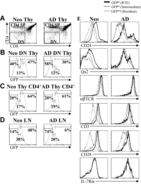
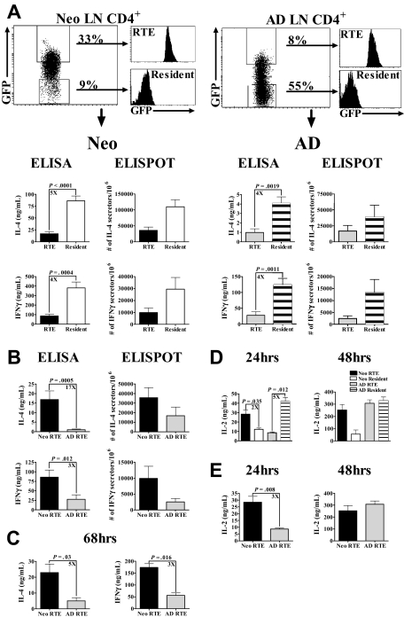
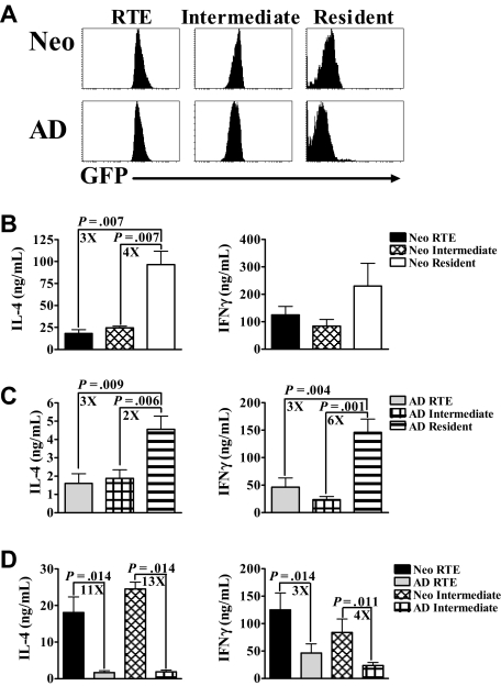
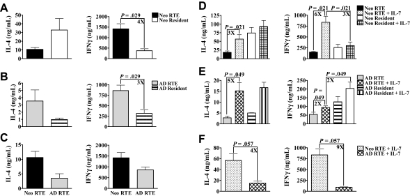
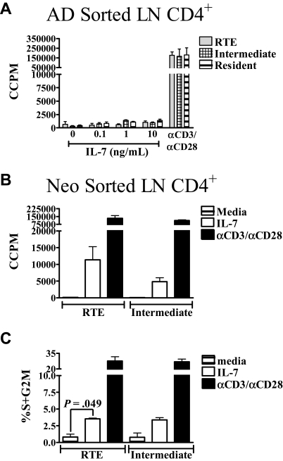
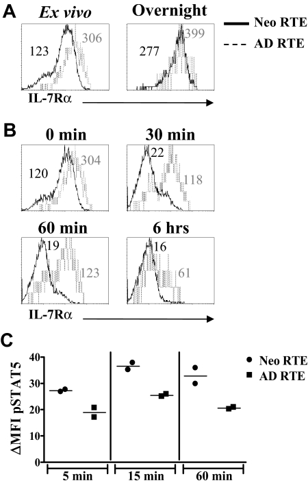
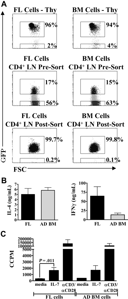
In contrast to adults, the murine neonatal CD4+ compartment contains a high frequency of recent thymic emigrants (RTEs). However, the functional capabilities of these cells in neonates are relatively unknown. Moreover, it has not been determined whether RTEs from neonates and adults are comparable. Here we have directly compared neonatal and adult CD4+ RTEs for the first time, using a transgenic mouse strain that allows for the identification and purification of RTEs. Our data demonstrate that RTEs from murine neonates and adults are phenotypically and functionally distinct. In particular, although the magnitude of RTEs cytokine responses from both age groups is dependent on the conditions of activation, neonatal RTEs always exhibited higher levels of effector Th1/Th2 cytokine production than adult RTEs. In addition, neonatal, but not adult, RTEs showed early proliferation in response to stimulation with interleukin-7 alone. This was associated with faster kinetics of interleukin-7Ralpha down-regulation and higher levels of pSTAT5 in neonatal RTEs. These quantitative and qualitative differences in the neonatal and adult RTEs populations may at least partially explain the diverse responses that are elicited in vivo in neonates in response to different conditions of antigen exposure.


Figures

Figure 1
Figure 1
RTEs are more abundant in neonates than adults. Thymocytes from neonatal (7-day-old) and adult (6- to 8-week-old) RAG2p-GFP+/− mice were stained for CD4 and CD8. (A) Gates were set on DN and CD4+ SP cells. (B) GFP expression within the gated DN thymocyte population was then used to define GFPhi RTEs, GFPlo intermediates, and GFP resident cells. The percentage of RTEs, intermediate, and resident cells among CD4+ SP thymocytes (C) and CD4+ LN cells (D) was then determined based on the gates set on DN thymocytes (B). (E) Using the GFP gates defined in panel B, the expression of CD24, Qa2, αβTCR, CD3, CD28, and IL-7Rα was determined on CD4+ RTEs, intermediate, and resident LN cells from neonates and adults. Histograms shown are representative of staining profiles from 6 to 14 individual neonates and 6 to 9 individual adults. For IL-7Rα, data represent 2 independent experiments, using a pool of LN cells from 14 or 15 neonates and 2 adults per experiment.
Figure 2
Figure 2
Neonatal RTEs produce higher levels of IL-2 and Th1/Th2 cytokines than adult RTEs in response to PBαCD3 and αCD28 stimulation. (A) GFPhi RTEs and GFP resident cells were sorted from purified neonatal and adult LN CD4+ cells, as described in “Preparation of cells from neonatal and adult RAG2p-GFP+/− mice.” Neonatal (left) and adult (right) RTEs and resident cells were activated for 48 hours with 0.5 μg/well PBαCD3 and 0.5 μg/mL soluble αCD28 (lower concentrations of PBαCD3, 0.005 or 0.05 μg/well, elicited only low-level cytokine production). Cytokine production and the number of cytokine-secreting cells were then determined. Graphs depict pooled data from 8 to 11 ELISA experiments and 3 ELISPOT experiments and are shown as the mean ± SEM; n = 35 to 60 neonates and 4 to 6 adults per experiment. (B) The data from panel A were regraphed to show a direct comparison of neonatal and adult RTEs. (C) Neonatal and adult RTEs were stimulated with PBαCD3 and αCD28 for 68 hours. Cytokine production was determined by ELISA. Graphs depict pooled data from 4 experiments; n = 18 to 35 neonates and 2 to 17 adults per experiment. (D) Sorted RTEs and resident cells from neonates and adults were stimulated with PBαCD3 and soluble αCD28 as described in panel A for 24 and 48 hours. Supernatants were harvested at each time point and assayed for IL-2 by ELISA. Graphs depict pooled data from 4 or 5 experiments; n = 18 to 25 neonates and 2 adults per experiment. (E) The data from panel D were regraphed to show a comparison of neonatal and adult RTEs.
Figure 3
Figure 3
Intermediate (GFPlo) cells from neonates also produce greater levels of cytokine compared with intermediate (GFPlo) cells from adults. (A) CD4+ LN cells from RAG2p-GFP+/− neonates and adults were sorted into RTEs (GFPhi), intermediate (GFPlo), and resident (GFP) populations. These purified neonatal (B) and adult (C) cells were then activated with PBαCD3 and αCD28 for 48 hours, and supernatants were harvested for cytokine-specific ELISA. (D) The data from panels B and C were regraphed to show a comparison of neonatal and adult RTEs and intermediate cells. Graphs represent pooled data from 4 or 5 independent experiments and are shown as the mean ± SEM; n = 35 to 60 neonates and 6 adults per experiment.
Figure 4
Figure 4
Relative response of RTEs and resident cells is dependent on the conditions of activation. (A-C) Sorted CD4+ LN RTEs and resident cells from RAG2p-GFP+/− neonates (A) and adults (B) were activated with PBαCD3 and APC for 48 hours. Supernatants were then harvested for cytokine-specific ELISA. (C) The data from panels A and B were regraphed to show a comparison of neonatal and adult RTEs activated with PBαCD3 and APC. Graphs represent pooled data from 4 independent experiments and are shown as the mean ± SEM; n = 34 to 44 neonates and 6 adults per experiment. (D-F) Sorted CD4+ LN RTEs and resident cells from neonatal (D) and adult (E) RAG2p-GFP+/− were activated with PBαCD3 and αCD28 in the presence or absence of 10 ng/mL IL-7 for 48 hours. Supernatants were then harvested for cytokine-specific ELISA. Graphs represent pooled data from 4 independent experiments and are shown as the mean ± SEM. (F) The data from panels D and E were regraphed to show a comparison of neonatal and adult RTEs activated in the presence of IL-7; n = 25 to 35 neonates; n = 4 to 6 adults per experiment.
Figure 5
Figure 5
Neonatal RTEs, but not adult RTEs, proliferate in response to IL-7. (A) CD4+ RTEs, intermediate, and resident LN cells were sorted from adults and cultured with increasing concentrations of IL-7 or PBαCD3 and αCD28 for 48 hours. 3H-thymidine was added during the last 20 hours of culture. (B) Neonatal RTEs and intermediate cells were sorted and cultured in complete media, 10 ng/mL IL-7, or activated with PBαCD3 and αCD28 for 48 hours. 3H-thymidine was added for the last 20 hours of culture. Graphs represent pooled data from 3 or 4 independent experiments; n = 6 to 8 adults and 28 to 44 neonates per experiment. All data are shown as the mean ± SEM. (C) CD4+ RTEs and intermediate cells were sorted from neonatal LN and then cultured in complete media, 10 ng/mL IL-7, or activated with PBαCD3 and αCD28 for 72 hours. The percentage of proliferating cells was determined by propidium iodide (PI) staining, as described in “Measurement of proliferation and cell cycle.” Graphs represent pooled data from 3 independent experiments; n = 23 to 56 neonates per experiment. All data are shown as the mean ± SEM.
Figure 6
Figure 6
IL-7Rα is down-regulated more rapidly on neonatal RTEs than on adult RTEs after exposure to IL-7 and is associated with higher levels of pSTAT5. (A) Purified CD4+ lymph node cells from RAG2p-GFP+/− neonates and adults were either stained directly ex vivo with anti–IL-7Rα (left panel) or were cultured overnight in complete media before staining (right panel). IL-7Rα expression on RTEs was determined by gating on CD4+GFPhi cells. (B) Purified CD4+ LN cells from neonatal and adult RAG2p-GFP+/− mice were either stained directly ex vivo (0 minutes) or cultured with 10 ng/mL IL-7 for 30 minutes, 60 minutes, or 6 hours, and then stained with anti–IL-7Rα. IL-7Rα expression on RTEs was determined by gating on CD4+GFPhi cells. IL-7Rα mean fluorescence intensity (MFI) for neonatal (in black) and adult (in gray) RTEs are shown in each histogram. Histograms represent data from 2 independent experiments; n = 2 adults and 15 neonates per experiment. In panels A and B, curves represent 800 to 2000 gated CD4+GFPhi RTEs for adults and 7000 to 13 000 gated CD4+GFPhi RTEs for neonates. (C) Sorted neonatal and adult RTEs were cultured overnight in complete media. The cells were then stimulated with IL-7 (10 ng/mL) for 5 to 60 minutes, fixed and permeabilized, and stained intracellularly for pSTAT5. The specificity of the staining was confirmed by competition with an excess of unlabeled pSTAT5 antibody (data not shown). Points represent pSTAT5 MFI data from 2 independent experiments; n = 40 to 49 neonates and 4 adults per experiment. ΔMFI was calculated by subtracting the MFI of pSTAT5 in the absence of IL-7 from the pSTAT5 MFI at each time point.
Figure 7
Figure 7
Functional characteristics of neonatal RTEs are not solely the result of the developmental age of the hematopoietic stem cell. Day 14 FL cells or adult BM cells from RAG2p-GFP+/− mice were transplanted intravenously into lethally irradiated wild-type RAG2p-GFP−/− adults. Six to 7 weeks later, LN CD4+ cells were purified and then sorted to obtain GFPhi RTEs. (A) Thymic reconstitution was similar in mice who received either FL (left panel) or BM cells (right panel). Both groups also possessed similar levels of GFPhi RTEs in the peripheral LN CD4+ compartment. Purity of sorted RTEs was > 99%. (B) Sorted RTEs were activated with PBαCD3 and αCD28 for 48 hours. Supernatants were then harvested for cytokine-specific ELISA. Data represent pooled data from 3 independent experiments and are shown as the mean ± SEM; n = 5 or 6 mice per group (FL and BM recipients). (C) Sorted RTEs were cultured in complete media or stimulated with either 10 ng/mL IL-7 or PBαCD3 and αCD28 for 48 hours. 3H-thymidine was added for the last 24 hours of culture to measure proliferation. Graphs represent pooled data from 3 independent experiments; n = 5 or 6 mice per group. All data are shown as the mean ± SEM.

Comment in

  • RTEs: lazy T-cell teenagers.
    Fink PJ. Fink PJ. Blood. 2009 May 28;113(22):5374-5. doi: 10.1182/blood-2009-03-207308. Blood. 2009. PMID: 19478052 No abstract available.

References

    1. Adkins B, Leclerc C, Marshall-Clarke S. Neonatal adaptive immunity comes of age. Nat Rev Immunol. 2004;4:553–564. - PubMed
    1. Scollay RG, Butcher EC, Weissman IL. Thymus cell migration: quantitative aspects of cellular traffic from the thymus to the periphery in mice. Eur J Immunol. 1980;10:210–218. - PubMed
    1. Boursalian TE, Golob J, Soper DM, Cooper CJ, Fink PJ. Continued maturation of thymic emigrants in the periphery. Nat Immunol. 2004;5:418–425. - PubMed
    1. Luettig B, Sponholz A, Heerwagen C, Bode U, Westermann J. Recent thymic emigrants (CD4+) continuously migrate through lymphoid organs: within the tissue they alter surface molecule expression. Scand J Immunol. 2001;53:563–571. - PubMed
    1. Bendelac A, Matzinger P, Seder RA, Paul WE, Schwartz RH. Activation events during thymic selection. J Exp Med. 1992;175:731–742. - PMC - PubMed

Publication types

MeSH terms