. 2009 Jun;50(6):2695-701.
doi: 10.1167/iovs.08-2919. Epub 2009 Jan 24.

Trichostatin a inhibits corneal haze in vitro and in vivo

Affiliations

Trichostatin a inhibits corneal haze in vitro and in vivo

Ajay Sharma et al. Invest Ophthalmol Vis Sci. 2009 Jun.

Abstract

Purpose: Trichostatin A (TSA), a histone deacetylase inhibitor, has been shown to suppress TGF-beta-induced fibrogenesis in many nonocular tissues. The authors evaluated TSA cytotoxicity and its antifibrogenic activity on TGF-beta-driven fibrosis in the cornea with the use of in vitro and in vivo models.

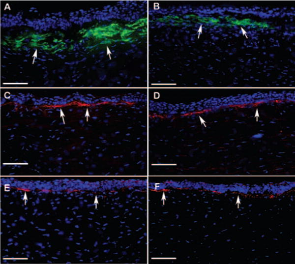
Methods: Human corneal fibroblasts (HSFs) were used for in vitro studies, and New Zealand White rabbits were used for in vivo studies. Haze in the rabbit cornea was produced with photorefractive keratectomy (PRK) using excimer laser. Trypan blue exclusion and MTT assays evaluated TSA cytotoxicity to the cornea. Density of haze in the rabbit eye was graded with slit lamp biomicroscopy. Real-time PCR, immunoblotting, or immunocytochemistry was used to measure alpha-smooth muscle actin (SMA), fibronectin, and collagen type IV mRNA or protein levels. TUNEL assay was used to detect cell death.

Results: TSA concentrations of 250 nM or less were noncytotoxic and did not alter normal HSF morphology or proliferation. TGF-beta1 treatment of HSF significantly increased mRNA and protein levels of SMA (9-fold), fibronectin (2.5-fold), and collagen type IV (2-fold). TSA treatment showed 60% to 75% decreases in TGF-beta1-induced SMA and fibronectin mRNA levels and 1.5- to 3.0-fold decreases in protein levels but had no effect on collagen type IV mRNA or protein levels in vitro. Two-minute topical treatment of TSA on rabbit corneas subjected to -9 D PRK significantly decreased corneal haze in vivo.

Conclusions: TSA inhibits TGF-beta1-induced accumulation of extracellular matrix and myofibroblast formation in the human cornea in vitro and markedly decreases haze in rabbit cornea in vivo.


Figures

FIGURE 1
FIGURE 1
Time- and dose-dependent cytotoxicity of TSA to cultured human corneal fibroblasts. Cultures were exposed to different concentrations of TSA (50, 100, 250, 500 nM) for 72 hours, and cell viability was quantified with MTT or trypan blue assay at 8, 24, 48, or 72 hours. TSA did not produce any cytotoxic effect on the cultured fibroblasts. Data are shown as mean ± SEM of percentage live cells. *P < 0.01 versus control.
FIGURE 2
FIGURE 2
Real-time quantitative PCR showing differential change in SMA (left), fibronectin (middle), and collagen type IV (right) expression in human corneal fibroblast cultures exposed to TGF-β1 (1 ng/mL) for 7 days under serum-free conditions with or without TSA. TGF-β1 caused a 9-fold increase in SMA, a 2.5-fold increase in fibronectin, and a 2-fold increase in collagen type IV levels. TSA treatment inhibited TGF-β1-induced SMA and fibronectin expression significantly. *P < 0.01 versus control; **P < 0.01 versus TGF-β1 treatment; ***P < 0.001 versus control.
FIGURE 3
FIGURE 3
Immunocytochemistry (left, middle) and immunoblotting (right) analyses showed significant decreases in myofibroblast formation by TSA in corneal fibroblast cultures grown in the presence of TGF-β1 (1 ng/mL) under serum-free conditions. Cell nuclei are stained blue with DAPI, and SMA-positive cells are stained green. Equal quantity of protein was loaded in each lane. GAPDH was used as a housekeeping gene. TSA treatment showed a significant 3-fold decrease in SMA and a 1.5-fold decrease in fibronectin expression. Scale bar, 100 μm.
FIGURE 4
FIGURE 4
(A) Representative slit lamp microscopy images demonstrating density and location of haze in control (left), PRK-treated (right), and PRK+0.02% TSA-treated (middle) rabbit corneas. (B) Biomicroscopic quantification of corneal haze. A significant decrease in corneal haze was observed in TSA-treated corneas (P < 0.01).
FIGURE 5
FIGURE 5
Representative immunohistochemistry images of corneal tissue sections showing levels of SMA (A, B), fibronectin (C, D), and collagen type IV (E, F) in rabbit corneas collected 4 weeks after −9 D PRK with or without TSA application. DAPI-stained nuclei are shown in blue, SMA-stained cells are shown in green, and fibronectin or collagen type IV-positive cells are shown in red. TSA-treated corneas showed significant decreases in SMA (B; P < 0.01) and notable decreases in fibronectin levels in the stroma compared with control corneas (A, C). No significant change was noted in collagen type IV (E, F). Arrows: positive protein immunostaining. Scale bar, 100 μm.
FIGURE 6
FIGURE 6
Quantification of SMA-positive cells/400 × column in the tissue section obtained from rabbit corneas that underwent −9 D PRK with or without topical application of 0.02% TSA. TSA treatment decreased SMA-positive cells by 60% (P < 0.01).
FIGURE 7
FIGURE 7
TUNEL assay of the central cornea from rabbits that underwent −9 D PRK with or without topical application of 0.02% TSA. Nuclei are stained blue with DAPI. Few TUNEL-positive cells (red) were seen in the epithelium. No loss of keratocytes (blue-stained nuclei) was noted in TSA-treated versus control (no TSA) rabbit corneas collected 4 weeks after −9 D PRK. Arrows: TUNEL-positive cells. Scale bar, 100 μm.

References

    1. Fong CS. Refractive surgery: the future of perfect vision? Singapore Med J. 2007;48:709–718. - PubMed
    1. Melki SA, Azar DT. LASIK complications: etiology, management, and prevention. Surv Ophthalmol. 2001;46:95–116. - PubMed
    1. McDonnell PJ. Emergence of refractive surgery. Arch Ophthalmol. 2000;118:1119–11120. - PubMed
    1. Shortt AJ, Bunce C, Allan BD. Evidence for superior efficacy and safety of LASIK over photorefractive keratectomy for correction of myopia. Ophthalmology. 2006;113:1897–1908. - PubMed
    1. Lee JB, Kim JS, Choe C, Seong GJ, Kim EK. Comparison of two procedures: photorefractive keratectomy versus laser in situ keratomileusis for low to moderate myopia. Jpn J Ophthalmol. 2001;45:487–491. - PubMed

Publication types

MeSH terms