. 2009 Feb;12(2):172-81.
doi: 10.1038/nn.2249. Epub 2009 Jan 25.

A critical role for PSD-95/AKAP interactions in endocytosis of synaptic AMPA receptors

Affiliations

A critical role for PSD-95/AKAP interactions in endocytosis of synaptic AMPA receptors

Samarjit Bhattacharyya et al. Nat Neurosci. 2009 Feb.

Abstract

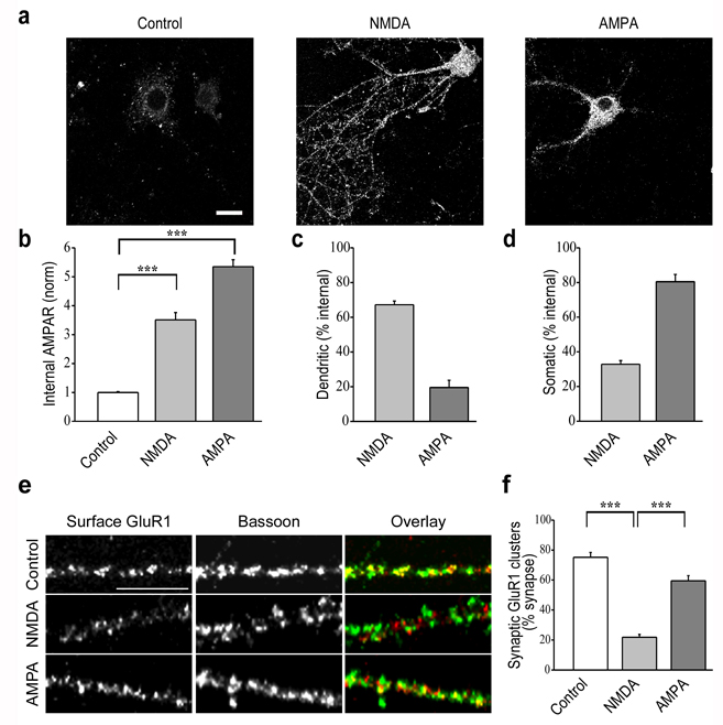
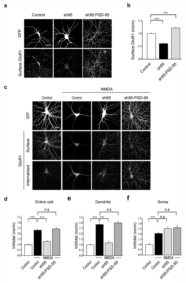
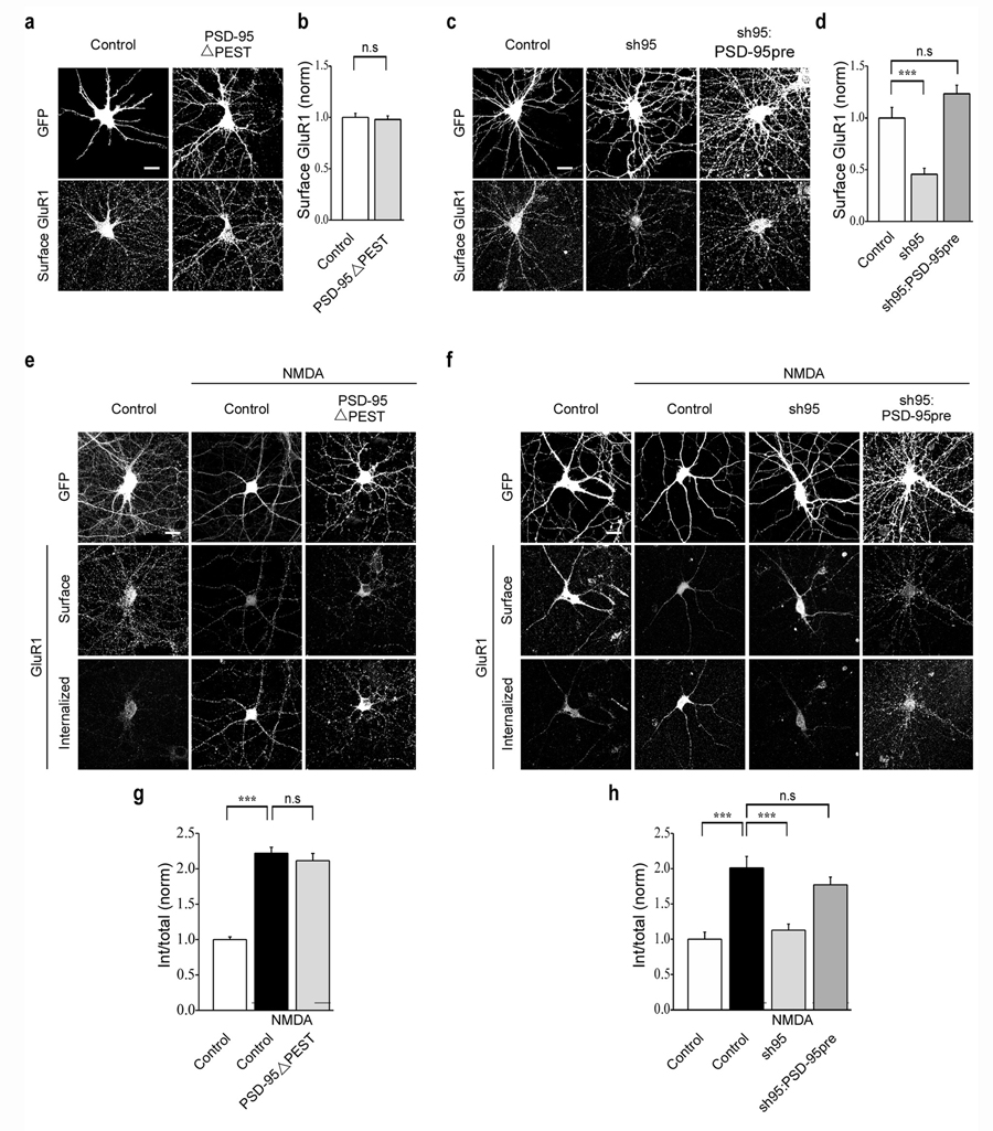
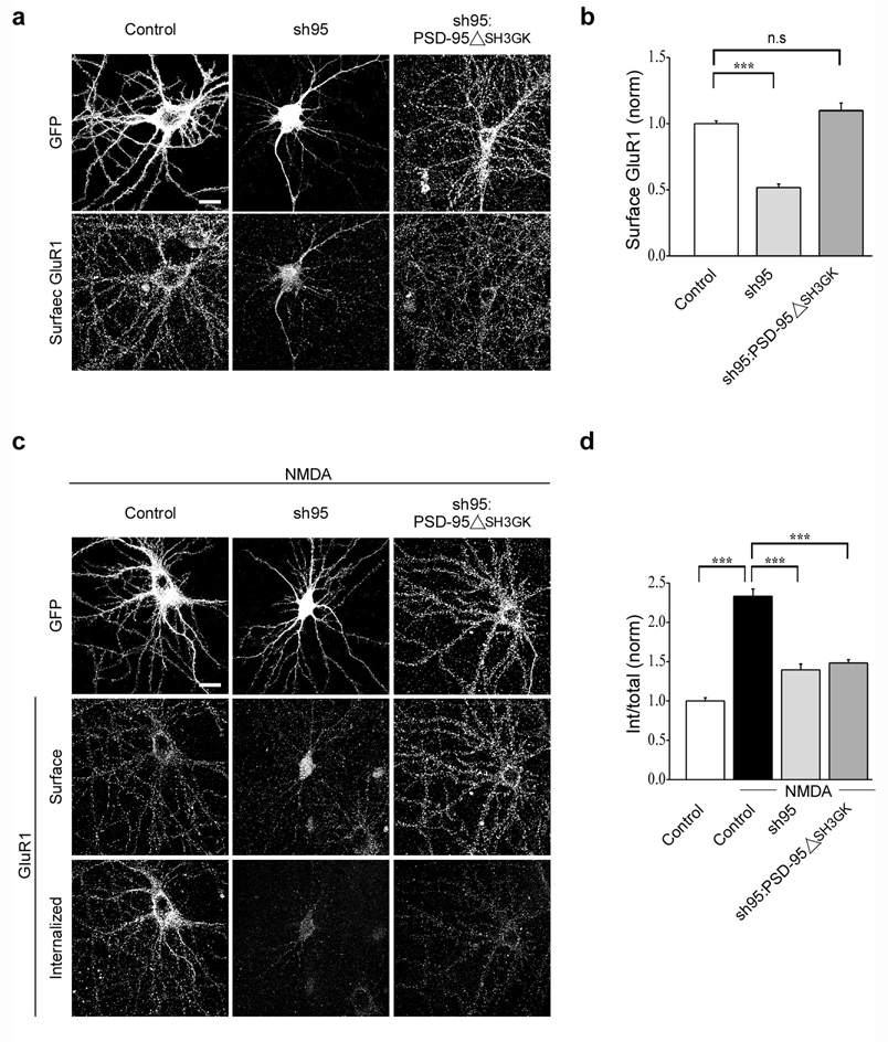
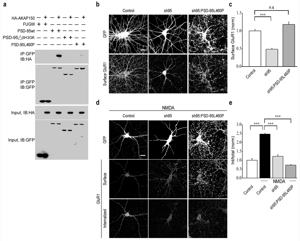
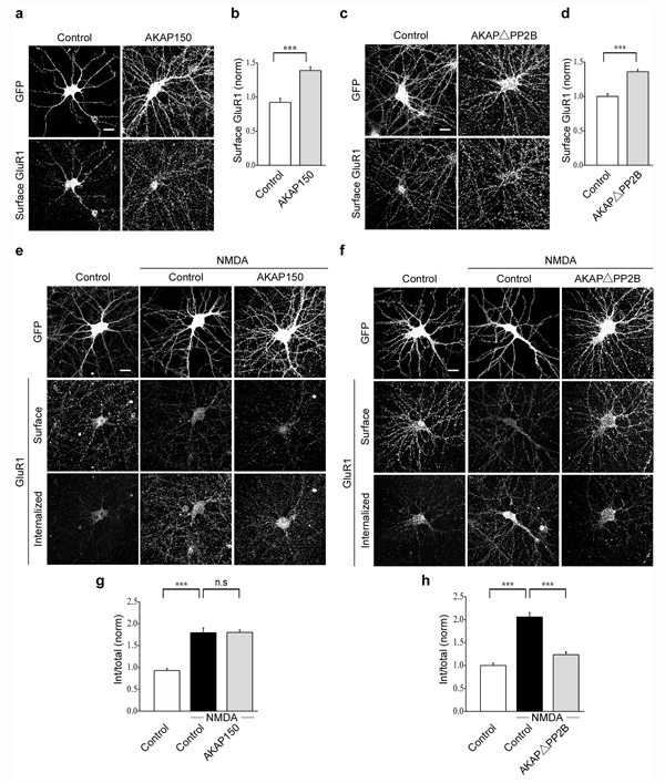
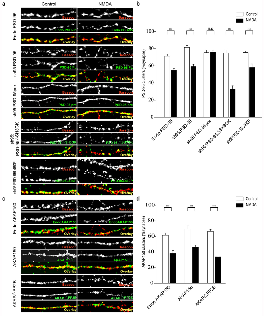
The endocytosis of AMPA receptors (AMPARs) underlies several forms of synaptic plasticity, including NMDA receptor (NMDAR)-dependent long-term depression (LTD), but the molecular mechanisms responsible for this trafficking remain unknown. We found that PSD-95, a major postsynaptic density protein, is important for NMDAR-triggered endocytosis of synaptic AMPARs in rat neuron cultures because of its binding to A kinase-anchoring protein 150 (AKAP150), a scaffold for specific protein kinases and phosphatases. Knockdown of PSD-95 with shRNA blocked NMDAR-triggered, but not constitutive or mGluR-triggered, endocytosis of AMPARs. Deletion of PSD-95's Src homology 3 and guanylate kinase-like domains, as well as a point mutation (L460P), both of which inhibit binding of PSD-95 to AKAP150, also blocked NMDAR-triggered AMPAR endocytosis. Furthermore, expression of a mutant AKAP150 that does not bind calcineurin inhibited this NMDAR-triggered trafficking event. Our results suggest that PSD-95's interaction with AKAP150 is critical for NMDAR-triggered AMPAR endocytosis and LTD, possibly because these scaffolds position calcineurin in the appropriate subsynaptic domain.


Figures

Figure 1
Figure 1
NMDAR and AMPAR-triggered endocytosis of AMPARs show distinct patterns of internalization. (a) Cells show internalized AMPAR immunoreactivity following NMDA treatment (100 µM for 3 min) in both distal dendrites and soma, whereas AMPA treatment (100 µM for 5 min) caused endocytosis of AMPARs primarily in proximal dendrites and the soma. Scale bar, 20 µm. (b) Quantitation of total endocytosis of AMPARs in response to different treatments. (c, d) Quantititation of dendritic (>10 µm from soma) versus somatic (soma and <10 µm from soma) AMPAR endocytosis. (e) Examples of dendritic staining for surface GluR1 (red) and Bassoon (green) after NMDA and AMPA treatment. Scale bar, 10 µm. (f) Quantitation of percent synapses (defined by Bassoon staining) that express detectable surface GluR1 puncta in control cells and cells that received NMDA or AMPA treatment. *** indicates p < 0.001 in all panels.
Figure 2
Figure 2
Acute knockdown of PSD-95 decreases the surface expression and NMDAR-triggered endocytosis of AMPARs. (a) Representative cells showing that surface GluR1 staining is reduced by sh95 compared to control cells expressing GFP and this reduction is rescued by expression of wildtype PSD-95 (fused to GFP). (b) Quantitation of surface GluR1 levels following knockdown and replacement of endogenous PSD-95. (c) Representative examples of surface and internalized AMPARs following NMDA treatment in cells expressing GFP, sh95 or sh95 and wildtype PSD-95. (d–f) Quantitation of the amount of NMDAR-triggered AMPAR endocytosis normalized to original surface levels throughout the cell (d), in the dendrites (e) and in the soma (f). Scale bar, 20 µm. *** indicates p < 0.001 and n.s indicates p > 0.05.
Figure 3
Figure 3
Constitutive endocytosis and mGluR-triggered endocytosis of AMPARs are not affected by knockdown of PSD-95. Representative images (a) and quantitation (c) of constitutive endocytosis of AMPARs in cells expressing GFP, sh95 or sh95 and wildtype PSD-95. Representative images (b) and quantitation (d) of AMPAR endocytosis triggered by DHPG application (50 µM for 5 min) in cells expressing GFP or sh95. (e) Bar graph showing that similar to NMDA application, DHPG application causes AMPAR endocytosis in both dendrites and soma. Scale bar, 20 µm. *** indicates p < 0.001 and n.s indicates p > 0.05.
Figure 4
Figure 4
PEST motif deletion and prenylation of PSD-95 does not affect NMDAR-triggered endocytosis of AMPARs. Representative images (a) and quantitation (b) of surface AMPAR (GluR1) staining in cells expressing GFP or PSD-95ΔPEST. Representative images (e) and quantitation (g) of NMDAR-triggered endocytosis of AMPARs in cells expressing GFP or PSD-95ΔPEST. Representative images (c) and quantitation (d) of surface AMPAR (GluR1) staining in cells expressing GFP, sh95 or sh95 and PSD-95pre. Note that the prenylated PSD-95 (PSD-95pre) rescues the decrease in AMPAR surface expression caused by sh95. Representative images (f) and quantitation (h) of NMDAR-triggered endocytosis of surface AMPARs in cells expressing GFP, sh95 or sh95 and PSD-95pre. Scale bar, 20 µm. *** indicates p < 0.001 and n.s indicates p > 0.05.
Figure 5
Figure 5
SH3 and GK domains of PSD-95 are required for NMDAR-triggered endocytosis of AMPARs. Representative images (a) and quantitation (b) of surface AMPARs in cells expressing GFP, sh95 or sh95 and PSD-95ΔSH3GK. Note that PSD-95ΔSH3GK rescues the surface expression of AMPARs. Representative images (c) and quantitation (d) of NMDAR-triggered endocytosis of surface AMPARs in cells expressing GFP, sh95 or sh95 and PSD-95ΔSH3GK. Scale bar, 20 µm. *** indicates p < 0.001 and n.s indicates p > 0.05.
Figure 6
Figure 6
L460P mutation in PSD-95 disrupts binding to AKAP150 and blocks NMDAR-triggered endocytosis of AMPARs. (a) Co-immunoprecipitation assays demonstrate that both deletion of SH3, GK domains and the L460P point mutation in full length PSD-95 disrupts binding of AKAP150 to PSD-95. Representative images (b) and quantitation (c) of surface AMPARs in cells expressing GFP, sh95 or sh95 and PSD-95L460P. Note that PSD-95L460P rescues the decrease in surface expression of AMPARs caused by sh95. Representative images (d) and quantitation (e) of NMDAR-triggered endocytosis of surface AMPARs in cells expressing GFP, sh95 or sh95 and PSD-95L460P. Scale bar, 20 µm. *** indicates p < 0.001 and n.s indicates p > 0.05.
Figure 7
Figure 7
Binding of calcineurin (PP2B) to AKAP150 is necessary for the NMDAR-triggered endocytosis of AMPARs. Overexpression of AKAP150 increases the surface expression of AMPARs as shown in the representative images (a) and quantitation (b) of surface AMPARs in cells expressing GFP or AKAP150. Representative images (e) and quantitation (g) of NMDAR-triggered endocytosis of surface AMPARs in cells expressing GFP or AKAP150. Representative images (c) and quantitation (d) of surface AMPARs in cells expressing GFP or AKAPΔPP2B. Representative images (f) and quantitation (h) of NMDAR-triggered endocytosis of surface AMPARs in cells expressing GFP or AKAPΔPP2B. scale bar, 20 µm. *** indicates p < 0.001 and n.s indicates p > 0.05.
Figure 8
Figure 8
NMDA application causes loss of PSD-95 and AKAP150 from synapses. Application of NMDA causes a decrease in the synaptic localization of endogenous PSD-95, recombinant wildtype PSD-95, PSD-95ΔSH3GK and PSD-95L460P but not prenylated PSD-95 as shown in the representative images (a) and quantitation (b) of PSD-95 puncta that co-localized with presynaptic Bassoon puncta. NMDA application also causes a loss of endogenous AKAP150, recombinant wildtype AKAP150 and AKAPΔPP2B from synapses as shown in the sample images (c) and quantitation (d). Scale bar, 10 µm. *** indicates p < 0.001 and n.s indicates p > 0.05.

References

    1. Malenka RC, Bear MF. LTP and LTD: an embarrassment of riches. Neuron. 2004;44:5–21. - PubMed
    1. Bredt DS, Nicoll RA. AMPA receptor trafficking at excitatory synapses. Neuron. 2003;40:361–379. - PubMed
    1. Collingridge GL, Isaac JT, Wang YT. Receptor trafficking and synaptic plasticity. Nat. Rev. Neurosci. 2004;5:952–962. - PubMed
    1. Shepherd JD, Huganir RL. The cell biology of synaptic plasticity: AMPA receptor trafficking. Annu. Rev. Cell Develop. Biol. 2007;23:613–643. - PubMed
    1. Hollmann M, Heinemann S. Cloned glutamate receptors. Annu. Rev. Neurosci. 1994;17:31–108. - PubMed

Publication types

MeSH terms