. 2009 Feb;16(2):176-82.
doi: 10.1038/nsmb.1546. Epub 2009 Jan 25.

Recognition of atypical 5' splice sites by shifted base-pairing to U1 snRNA

Affiliations

Recognition of atypical 5' splice sites by shifted base-pairing to U1 snRNA

Xavier Roca et al. Nat Struct Mol Biol. 2009 Feb.

Abstract

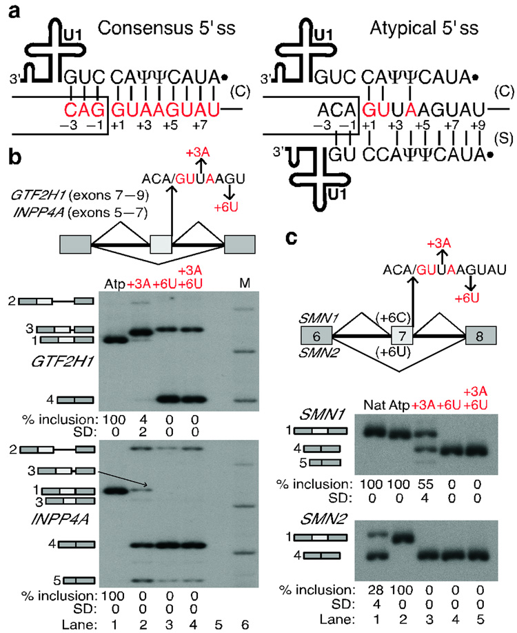
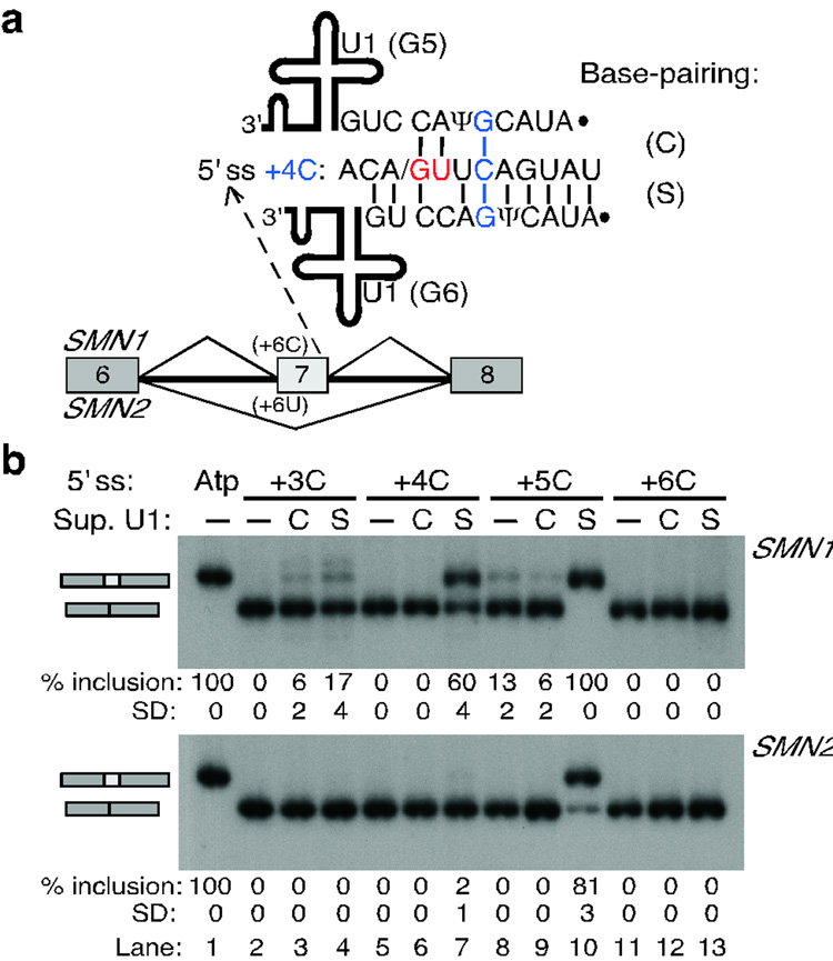
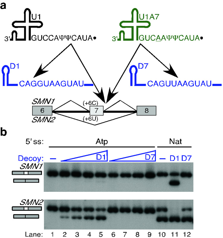
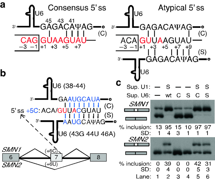
Accurate pre-mRNA splicing is crucial for gene expression. The 5' splice site (5' ss)--the highly diverse element at the 5' end of introns--is initially recognized via base-pairing to the 5' end of the U1 small nuclear RNA (snRNA). However, many natural 5' ss have a poor match to the consensus sequence, and are predicted to be weak. Using genetic suppression experiments in human cells, we demonstrate that some atypical 5' ss are actually efficiently recognized by U1, in an alternative base-pairing register that is shifted by one nucleotide. These atypical 5' ss are phylogenetically widespread, and many of them are conserved. Moreover, shifted base-pairing provides an explanation for the effect of a 5' ss mutation associated with pontocerebellar hypoplasia. The unexpected flexibility in 5' ss-U1 base-pairing challenges an established paradigm and has broad implications for splice-site prediction algorithms and gene-annotation efforts in genome projects.


Figures

Figure 1
Figure 1. Shifted base-pairing between atypical 5' ss and the 5' end of U1 snRNA
a, Diagram of the two base-pairing registers between the 5' ss (positions are numbered) and U1. Consensus nucleotides are shown in red in all figures (see Methods). Ψ, pseudouridine. Solid dot, 2,2,7-trimethylguanosine cap at the 5' end of U1. Box, upstream exon; line, intron. Base pairs in the canonical (C) or shifted (S) register are indicated by vertical lines. Note that the atypical 5' ss can form seven more base pairs to U1 in the shifted arrangement. b, Mutations at atypical (Atp) 5' ss that disrupt shifted but enhance canonical base-pairing abolish correct splicing. The human GTF2H1 and INPP4A minigenes are schematically represented at the top, indicating the mutations introduced at the atypical 5' ss. M, Molecular weight markers. The identity of the various spliced mRNAs, detected by radioactive RT-PCR, is schematically shown on the left of the gels and corresponds to: #1, correctly spliced mRNA; #2, retention of the downstream intron; #3, use of cryptic 5' ss in the middle exon; #4, skipping of the middle exon; #5, activation of a cryptic 5' ss in the first exon. The percentage of correct splicing is shown at the bottom. See Supplementary Fig. 1 online for details about the aberrantly spliced mRNAs. c, RT-PCR analysis of the atypical 5' ss in the SMN1/2 context (schematic at the top). Nat, natural SMN1/2 exon 7 5' ss. Numbers below the panels show the percentage and Standard Deviation (SD) of exon 7 inclusion.
Figure 2
Figure 2. Suppressor U1 snRNAs in the shifted register can rescue splicing
a, Schematic of the single mutations introduced at the atypical 5' ss in the SMN1/2 context. These mutations substitute a non-consensus nucleotide by a consensus nucleotide. b, Base-pairing of the mutant 5' ss with the corresponding suppressor U1 snRNA. As an example, we show the base-pairing of the +5G mutant 5' ss with the suppressor U1 snRNA carrying the corresponding compensatory mutation (C5) in the shifted register. The mutant nucleotide at the 5' end of U1 in each case is shown in red. See Supplementary Fig. 2 online for the base-pairing of all mutant 5' ss with their respective suppressor U1s. c, RT-PCR analysis of the SMN1/2 minigenes carrying the wild-type (lane 1) or mutant atypical 5' ss (lanes 2–13). The 5' ss mutation is indicated at the top, without (−) or with (+) the corresponding suppressor U1 snRNA. The mRNA products are schematically indicated on the left of each panel. The fastest migrating band in SMN1 corresponds to an mRNA that skipped exon 7 and used a cryptic 5' ss 50 nucleotides upstream of the exon 6 5' ss. The percentage and Standard Deviation (SD) of exon 7 inclusion is indicated below each autoradiogram.
Figure 3
Figure 3. Compensatory U1 mutations that restore shifted but not canonical base-pairing rescue splicing at atypical 5' ss
a, Scheme of the experimental design. SMN1/2 minigenes carrying point mutations at a heterologous atypical 5' ss in exon 7 were co-transfected with suppressor U1 snRNAs. The 5' ss nucleotides at positions +3 to +6 were individually mutated to C (+3C to +6C). The 5' end of U1 was mutated so as to rescue base-pairing in the canonical or the shifted arrangement (suppressor U1 mutations G3 to G6). Mutant +4C is shown as a representative example, for which U1 mutations G5 or G6 restore base-pairing in the canonical (C) or shifted (S) register, respectively. For the other three mutations, see Supplementary Fig. 2 online. b, RT-PCR analysis of the +3C to +6C 5' ss mutations in SMN1/2 with suppressor U1. Top labels indicate the 5' ss mutant and the suppressor U1 in either register. Atp, wild-type atypical 5' ss. The percentage and Standard Deviation of exon 7 inclusion is shown below each autoradiogram.
Figure 4
Figure 4. U1 but not U1A7 snRNA decoys reduce splicing via the atypical 5' ss
a, Schematic of the U1 (black) and U1A7 (green) snRNA decoys. The D1 and D7 decoys are short RNAs expressed from the potent U6 promoter, and comprise the first 27 nucleotides of the U6 snRNA for stability, and a sequence with perfect complementarity to the 5' end of U1 (black) or U1A7 (green) snRNAs, respectively. These decoys reduce the free levels of their cognate snRNAs in the cell, affecting the splicing of certain introns. b, The D1 but not the D7 decoy reduced SMN1/2 exon 7 inclusion in minigenes carrying the natural or an atypical 5' ss. The top labels indicate the identity of the 5' ss in exon 7 and the decoy used. The triangle depicts an increasing amount of decoy plasmid transfected with the minigene.
Figure 5
Figure 5. U6 snRNA does not base-pair to the atypical 5' ss in a shifted register
a, Schematic of the base-pairing between consensus (left) or atypical (right) 5' ss and the conserved U6 ACAGAG box (positions are numbered). The open dot indicates the γ-monomethyl cap. The atypical 5' ss has an extended base-pairing potential to U6 in the shifted register. b, Schematic of the suppressor U6 snRNAs carrying compensatory mutations in either the canonical (C) or the shifted (S) register. These mutations (blue font) restore base-pairing for the +5C mutation at atypical 5' ss in the SMN1/2 context. c, RT-PCR analysis of the SMN1/2 minigenes cotransfected with suppressor U1 and U6 snRNAs. Top labels indicate the suppressor U1/U6 used. wt, wild-type U6 snRNA. Suppressor U6s alone had no effect (lanes 3, 4 vs. lane 1). In combination with suppressor U1, suppressor U6 in the canonical register resulted in more exon 7 inclusion than suppressor U6 in the shifted register (lanes 5 and 6 in SMN2). These results suggest that atypical 5' ss establish canonical base-pairing to U6 snRNA.

References

    1. Brow DA. Allosteric cascade of spliceosome activation. Annu. Rev. Genet. 2002;36:333–360. - PubMed
    1. Bessonov S, Anokhina M, Will CL, Urlaub H, Lührmann R. Isolation of an active step I spliceosome and composition of its RNP core. Nature. 2008;452:846–850. - PubMed
    1. Lerner MR, Boyle JA, Mount SM, Wolin SL, Steitz JA. Are snRNPs involved in splicing? Nature. 1980;283:220–224. - PubMed
    1. Rogers J, Wall R. A mechanism for RNA splicing. Proc. Natl. Acad. Sci. USA. 1980;77:1877–1879. - PMC - PubMed
    1. Zhuang Y, Weiner AM. A compensatory base change in U1 snRNA suppresses a 5' splice site mutation. Cell. 1986;46:827–835. - PubMed

Publication types