

Electrolyte and Fluid Transport in Mesothelial Cells
- PMID: 19169368
- PMCID: PMC2629602
- DOI: 10.2174/1875044300801010001
Electrolyte and Fluid Transport in Mesothelial Cells
Abstract
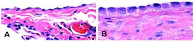
Mesothelial cells are specialized epithelial cells, which line the pleural, pericardial, and peritoneal cavities. Accumulating evidence suggests that the monolayer of mesothelial cells is permeable to electrolyte and fluid, and thereby govern both fluid secretion and re-absorption in the serosal cavities. Disorders in these salt and fluid transport systems may be fundamental in the pathogenesis of pleural effusion, pericardial effusion, and ascites. In this review, we discuss the location, physiological function, and regulation of active transport (Na(+)-K(+)-ATPase) systems, cation and anion channels (Na(+), K(+), Cl(-), and Ca(2+) channels), antiport (exchangers) systems, and symport (co-transporters) systems, and water channels (aquaporins). These secretive and absorptive pathways across mesothelial monolayer cells for electrolytes and fluid may provide pivotal therapeutical targets for novel clinical intervention in edematous diseases of serous cavities.
Figures

Similar articles
-
Regulation of Cl- Electrolyte Permeability in Epithelia by Active Traditional Chinese Medicine Monomers for Diarrhea.Curr Drug Targets. 2020;21(9):902-909. doi: 10.2174/1389450121666200504073635. Curr Drug Targets. 2020. PMID: 32364074 Review.
-
Regulation of Electrolyte Permeability by Herbal Monomers in Edematous Disorders.Curr Pharm Des. 2021;27(6):833-839. doi: 10.2174/1381612826666200917144655. Curr Pharm Des. 2021. PMID: 32940173 Review.
-
Tubular fluid secretion in the seminiferous epithelium: ion transporters and aquaporins in Sertoli cells.J Membr Biol. 2010 Jul;236(2):215-24. doi: 10.1007/s00232-010-9294-x. Epub 2010 Aug 10. J Membr Biol. 2010. PMID: 20697886 Review.
-
Transepithelial Fluid and Salt Re-Absorption Regulated by cGK2 Signals.Int J Mol Sci. 2018 Mar 16;19(3):881. doi: 10.3390/ijms19030881. Int J Mol Sci. 2018. PMID: 29547542 Free PMC article. Review.
-
Basolateral ion transporters involved in colonic epithelial electrolyte absorption, anion secretion and cellular homeostasis.Acta Physiol (Oxf). 2011 Jan;201(1):33-46. doi: 10.1111/j.1748-1716.2010.02153.x. Acta Physiol (Oxf). 2011. PMID: 20528802 Review.
Cited by
-
Multiple cell types in the oviduct express the prolactin receptor.FASEB Bioadv. 2022 Apr 15;4(7):485-504. doi: 10.1096/fba.2022-00004. eCollection 2022 Jul. FASEB Bioadv. 2022. PMID: 35812077 Free PMC article.
-
δ ENaC: a novel divergent amiloride-inhibitable sodium channel.Am J Physiol Lung Cell Mol Physiol. 2012 Dec 15;303(12):L1013-26. doi: 10.1152/ajplung.00206.2012. Epub 2012 Sep 14. Am J Physiol Lung Cell Mol Physiol. 2012. PMID: 22983350 Free PMC article. Review.
-
Effusion-Serum Chloride Gradient in Heart Failure-Associated Pleural Effusion - Pathophysiologic Implications.Circ Rep. 2020 Jun 2;2(7):357-363. doi: 10.1253/circrep.CR-20-0018. Circ Rep. 2020. PMID: 33693253 Free PMC article.
-
Aquaporin-1 Facilitates Transmesothelial Water Permeability: In Vitro and Ex Vivo Evidence and Possible Implications in Peritoneal Dialysis.Int J Mol Sci. 2021 Nov 21;22(22):12535. doi: 10.3390/ijms222212535. Int J Mol Sci. 2021. PMID: 34830416 Free PMC article.
-
Expression and regulation of epithelial Na+ channels by nucleotides in pleural mesothelial cells.Am J Respir Cell Mol Biol. 2009 May;40(5):543-54. doi: 10.1165/rcmb.2008-0166OC. Epub 2008 Oct 16. Am J Respir Cell Mol Biol. 2009. PMID: 18927349 Free PMC article.
References
-
- Lai-Fook SJ. Pleural mechanics and fluid exchange. Physiol Rev. 2004 Apr;84:385–410. - PubMed
-
- Yung S, Li FK, Chan TM. Peritoneal mesothelial cell culture and biology. Perit Dial Int. 2006 Mar–Apr;26:162–73. - PubMed
-
- Zocchi L. Physiology and pathophysiology of pleural fluid turnover. Eur Respir J. 2002 Dec;20:1545–58. - PubMed
-
- Canessa CM, Schild L, Buell G, Thorens B, Gautschi I, Horisberger JD, Rossier BC. Amiloride-sensitive epithelial Na+ channel is made of three homologous subunits. Nature. 1994 Feb 3;367:463–7. - PubMed
-
- Lingueglia E, Voilley N, Waldmann R, Lazdunski M, Barbry P. Expression cloning of an epithelial amiloride-sensitive Na+ channel. A new channel type with homologies to Caenorhabditis elegans degenerins. FEBS Lett. 1993 Feb 22;318:95–9. - PubMed
Grants and funding
LinkOut - more resources
Full Text Sources
Miscellaneous