

doi: 10.1016/j.cnr.2006.09.004.
The cellular response in neuroinflammation: The role of leukocytes, microglia and astrocytes in neuronal death and survival
Affiliations
- PMID: 19169437
- PMCID: PMC2630233
- DOI: 10.1016/j.cnr.2006.09.004
Item in Clipboard
The cellular response in neuroinflammation: The role of leukocytes, microglia and astrocytes in neuronal death and survival
Clin Neurosci Res.
2006 Dec.
Abstract
Neuroinflammation is a complex integration of the responses of all cells present within the CNS, including the neurons, macroglia, microglia and the infiltrating leukocytes. The initiating insult, environmental factors, genetic background and age/past experiences all combine to modulate the integrated response of this complex neuroinflammatory circuit. Here, we explore how these factors interact to lead to either neuroprotective versus neurotoxic inflammatory responses. We specifically focus on microglia and astrocytic regulation of autoreactive T cell responses.
Figures
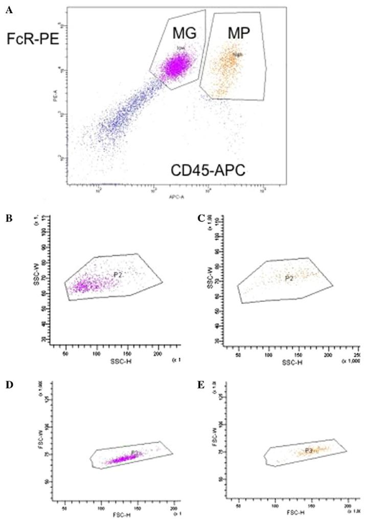
Flow cytometric analysis of microglia and macrophages isolated from LPS injected murine CNS. (A) Microglia (MG) are defined as FcR+, CD45 low cells, macrophages (MP) are defined as FcR+, CD45 high cells. (B) depicts the side scatter (SSC-H, SSC-W) of just the microglia defined in (A). (C) Depicts the side scatter (SSC-H, SSC-W) of just the macrophages defined in (A). (D) Depicts the forward scatter (FSC-H, FSC-W) of just the microglia defined in (A). (E) depicts the forward scatter (FSC-H, FSC-W) of just the macrophages defined in (A).
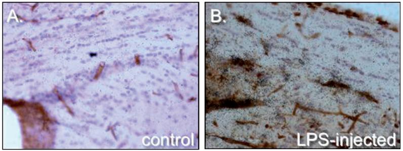
Induction of ceruloplasmin in the corpus callosum by intracerebral injection of LPS. Brain sections depict ceruloplasmin expression in healthy adult mouse brain (A) and 24 h following LPS injection (B). Ceruloplasmin expression is visualized by 33P labeled riboprobes (dark emulsion grains), cell nuclei are visualized by hematoxylin (blue) and areas of inflammation are visualized by increased labeling of microglia, macrophages and blood vessels with tomato lectin (in brown).
References
-
- Lo D, Feng LL, Li L, Carson MJ, Crowley M, Pauza M, et al. Integrating innate and adaptive immunity in the whole animal. Immunol Rev. 1999;169:225–39. - PubMed
-
- Medzhitov R, Janeway CA., Jr Innate immune recognition and control of adaptive immune responses. Semin Immunol. 1998;10(5):351–3. - PubMed
-
- Bechmann I. Failed central nervous system regeneration: a downside of immune privilege? Neuromol Med. 2005;7(3):217–28. - PubMed
Grants and funding
LinkOut - more resources
Full Text Sources
Other Literature Sources
Medical
Miscellaneous