. 2009 Jun;219(3):584-94.
doi: 10.1002/jcp.21699.

Phytoestrogens regulate mRNA and protein levels of guanine nucleotide-binding protein, beta-1 subunit (GNB1) in MCF-7 cells

Affiliations

Phytoestrogens regulate mRNA and protein levels of guanine nucleotide-binding protein, beta-1 subunit (GNB1) in MCF-7 cells

Srivatcha Naragoni et al. J Cell Physiol. 2009 Jun.

Abstract

Phytoestrogens (PEs) are non-steroidal ligands, which regulate the expression of number of estrogen receptor-dependent genes responsible for a variety of biological processes. Deciphering the molecular mechanism of action of these compounds is of great importance because it would increase our understanding of the role(s) these bioactive chemicals play in prevention and treatment of estrogen-based diseases. In this study, we applied suppression subtractive hybridization (SSH) to identify genes that are regulated by PEs through either the classic nuclear-based estrogen receptor or membrane-based estrogen receptor pathways. SSH, using mRNA from genistein (GE) treated MCF-7 cells as testers, resulted in a significant increase in GNB1 mRNA expression levels as compared with 10 nM 17beta estradiol or the no treatment control. GNB1 mRNA expression was up regulated two- to fivefold following exposure to 100.0 nM GE. Similarly, GNB1 protein expression was up regulated 12- to 14-fold. GE regulation of GNB1 was estrogen receptor-dependent, in the presence of the anti-estrogen ICI-182,780, both GNB1 mRNA and protein expression were inhibited. Analysis of the GNB1 promoter using ChIP assay showed a PE-dependent association of estrogen receptor alpha (ERalpha) and beta (ERbeta) to the GNB1 promoter. This association was specific for ERalpha since association was not observed when the cells were co-incubated with GE and the ERalpha antagonist, ICI. Our data demonstrate that the levels of G-protein, beta-1 subunit are regulated by PEs through an estrogen receptor pathway and further suggest that PEs may control the ratio of alpha-subunit to beta/gamma-subunits of the G-protein complex in cells. J. Cell. Physiol. 219: 584-594, 2009. (c) 2009 Wiley-Liss, Inc.


Figures

Figure I
Figure I. Identification of differentially expressed sequences by suppression subtractive hybridization
MCF-7 cells were grown in the presence and absence of 100 nM GE or 10 nM 17-β E2 for 24 h. DNase I-treated mRNA obtained from these cells was subjected to PCR-select subtractive hybridization as described in Materials and Methods. (A) pS2 expression following exposure to GE or 17-β E2. (B) Agarose gel of differential PCR products (DP) obtained after subtractive hybridization. Lane 1, DP1 (control verses GE); Lane 2, DP2 (DP1 verses 17-β E2); Lane 3 amplification of DP2 with nested PCR primers; Lane 4, SSH Control, human skeletal muscle cDNA spiked with plasmid DNA as tester; Lane 5, amplification of SSH control with nested PCR primer. (C) PCR analysis of subtraction efficiency. PCR was performed on driver (CON or 17-β E2), unsubtracted tester (GE), and subtracted product (DP2) using GAPDH, pS2 or PgR specific primers.
Figure II
Figure II. Quantification of differentially expressed genes via reverse Northern blot analysis
Differential products (DP2) obtained from SSH between DP1 and 17-β E2 were cloned into a pGEM-T cloning vector and several individual clones were dot-blotted onto nylon membranes. GE differential expression clones (A) or 17-β E2 differential expression clones (B) were identified by probing the blot with equal amounts of 32P cDNA prepared from reverse transcribed RNA obtained from MCF-7 cells treated with 100 nM GE or 10 nM 17-β E2 as described in Materials and Methods. Circles represent those genes that are regulated by GE, stars represent genes that are regulated by 17-β E2 and squares represent genes that show dual regulation by both GE and 17-β E2. (C) Fold expression of GE responsive genes relative to 17-β E2. The signal on the autoradiograph in blot A and blot B was quantified using the OptiQuant Image analysis software and the digital light units in blot A (GE) were normalized to that in blot B (17-β E2).
Figure III
Figure III. Differential regulation of GNB1 gene by Genistein in MCF-7 cells.
(A) Relative quantification of GNB1 mRNA expression. Total RNA samples from GE treated MCF-7 cells were analyzed using SYBR Green Real Time RT-PCR. Relative quantification of GNB1 mRNA (Insert) was performed against the endogenous GAPDH gene and normalized with the no treatment control RNA. NT, no treatment control sample, NTC, no template control for PCR (B) Quantification of GNB1 in DP2 library. Plasmid DNA (1 ng) isolated from the DP2 library was amplified by SYBR Green Real Time PCR using gene specific primers for GNB1, pS2, PgR, and GAPDH. (C) GNB1 protein expression in MCF-7. MCF-7 cells were grown in 100 nM GE plus or minus ICI for 24 h, and 50 μg of total protein was analyzed by Western blot using anti-GNB1 primary antibody. The blot was probed with anti-GAPDH which was used as a loading control. (D). Northern blot analysis of GE induced GNB1 expression. Total RNA (30 μg) from GE treated MCF-7 was isolated and used in Northern blot analysis with 32P-labeled GNB1 cDNA as probe. The 18s rRNA was used as a loading control for Northern blot analysis.
Figure IV
Figure IV. Up regulation of GNB1 mRNA by phytoestrogen
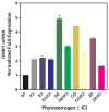
Total RNA (30 μg) was isolated from MCF-7 cells treated with 17-β E2 (10 nM) or PE (100 nM) ± 1 μM ICI antiestrogen for 24 h. Samples were separated by formaldehyde-agarose gel electrophoresis, transferred to a nylon membrane, UV cross-linked, and hybridized with 32P-labeled GNB1 cDNA probe generated by restriction enzyme digestion. The hybridized signals were scanned on a Packard Cyclone PhosphoImager and quantified using the OptiQuant Imager analysis software. Relative quantification of GNB1 mRNA expression was performed against the loading control 18s rRNA and normalized with the no treatment control. NT, no treatment control; ICI, antiestrogen ICI 182,780; 17-β Estradiol; GE, Genistein; CO, Coumestrol; and ZE, Zearalenone.
Figure V
Figure V. Phytoestrogen regulatation of GNB1 protein expression through both ERα and ERβ
MCF-7 cells were induced with GE in the presence and absence of ERα{antagonist (ICI) and ERβ antagonist (R,R-THC). Cellular GNB1 levels were determined by western blot analysis using anti-GNB1 antibody. Immunoreactive bands were visualized using enhanced chemilluminescence and the resulting signal was quantified with the AlphaEaseFC software. Protein expression of GAPDH was used as an internal control. Fold expression of GNB1 was normalized to the no-treatment control sample.
Figure VI
Figure VI. Phytoestrogen-induced association of ERα with the GNB1 promoter
MCF-7 cells were induced with 100 nM phytoestrogens (genistein and coumestrol) in the presence and absence of 1 μM ICI for 24 h. The cells were cross-linked with 1% formaldehyde and the cross-linked DNA-protein complex fragmented by sonication followed by immunoprecipitation with anti-ERα antibody. The immunoprecipitated chromatin was analyzed by PCR for GNB1 promoter DNA and by Western for ERα and ERβ proteins. (A) Schematic representation of GNB1 indicating an ERE/Sp 1 site and the region amplified by GNB1 promoter primers. (B) PCR of GNB1 promoter immunoprecipitated by ERα antibody (left) and Western blot of immunoprecipitated protein (right). (C) PCR amplification of DNA from IP input (left) and PCR amplification of IP using primer pairs that amplify a region of GNB1 that does not contain ERE/Sp 1 binding sites (right), establishing that phytoestrogen recruits ERα to the GNB1 gene. (D) PCR of GNB1 promoter immunoprecipitated by ERβ antibody (left) and Western blot of immunoprecipitated protein (right).

Similar articles

Cited by

References

    1. Adlercreutz H, Bannwart C, Wahala K, Makela T, Brunow G, Hase T, Arosemena PJ, Kellis JT, Jr, Vickery LE. Inhibition of human aromatase by mammalian lignans and isoflavonoid phytoestrogens. J Steroid Biochem Mol Biol. 1993;44(2):147–153. - PubMed
    1. Ahn CH, Jeong EG, Lee JW, Kim MS, Kim SH, Kim SS, Yoo NJ, Lee SH. Expression of beclin-1, an autophagy-related protein, in gastric and colorectal cancers. Apmis. 2007;115(12):1344–1349. - PubMed
    1. Allred CD, Ju YH, Allred KF, Chang J, Helferich WG. Dietary genistin stimulates growth of estrogen-dependent breast cancer tumors similar to that observed with genistein. Carcinogenesis. 2001;22(10):1667–1673. - PubMed
    1. Altschul SF, Madden TL, Schaffer AA, Zhang J, Zhang Z, Miller W, Lipman DJ. Gapped BLAST and PSI-BLAST: a new generation of protein database search programs. Nucleic Acids Res. 1997;25(17):3389–3402. - PMC - PubMed
    1. Ansonoff MA, Etgen AM. βββ elevates protein kinase C catalytic activity in the preoptic area of female rats. Endocrinology. 1998;139(7):3050–3056. - PubMed

MeSH terms

LinkOut - more resources