Noninvasive evaluation of oral lesions using depth-sensitive optical spectroscopy
- PMID: 19170229
- PMCID: PMC2728679
- DOI: 10.1002/cncr.24177
Noninvasive evaluation of oral lesions using depth-sensitive optical spectroscopy
Abstract
Background: Optical spectroscopy is a noninvasive technique with potential applications for diagnosis of oral dysplasia and early cancer. In this study, we evaluated the diagnostic performance of a depth-sensitive optical spectroscopy (DSOS) system for distinguishing dysplasia and carcinoma from non-neoplastic oral mucosa.
Methods: Patients with oral lesions and volunteers without any oral abnormalities were recruited to participate. Autofluorescence and diffuse reflectance spectra of selected oral sites were measured using the DSOS system. A total of 424 oral sites in 124 subjects were measured and analyzed, including 154 sites in 60 patients with oral lesions and 270 sites in 64 normal volunteers. Measured optical spectra were used to develop computer-based algorithms to identify the presence of dysplasia or cancer. Sensitivity and specificity were calculated using a gold standard of histopathology for patient sites and clinical impression for normal volunteer sites.
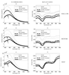
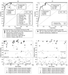
Results: Differences in oral spectra were observed in: (1) neoplastic versus nonneoplastic sites, (2) keratinized versus nonkeratinized tissue, and (3) shallow versus deep depths within oral tissue. Algorithms based on spectra from 310 nonkeratinized anatomic sites (buccal, tongue, floor of mouth, and lip) yielded an area under the receiver operating characteristic curve of 0.96 in the training set and 0.93 in the validation set.
Conclusions: The ability to selectively target epithelial and shallow stromal depth regions appeared to be diagnostically useful. For nonkeratinized oral sites, the sensitivity and specificity of this objective diagnostic technique were comparable to that of clinical diagnosis by expert observers. Thus, DSOS has potential to augment oral cancer screening efforts in community settings.
Figures


References
-
- Stewart BW, Kleihues P, editors. World Cancer Report. Lyon: IARCPress; 2003.
-
- Ries LAG, Melbert D, Krapcho M, et al., editors. SEER Cancer Statistics Review, 1975–2005, National Cancer Institute. http://seer.cancer.gov/csr/1975_2005/, based on November 2007 SEER data submission, posted to the SEER web site. 2008.
-
- Neville BW, Day TA. Oral cancer and precancerous lesions. CA Cancer J Clin. 2002;52:195–215. - PubMed
-
- Chen AY, Myers JN. Cancer of the oral cavity. Dis Mon. 2001;47:265–361. - PubMed
-
- Fryen A, Glanz H, Lohmann W, Dreyer T, Bohle RM. Significance of autofluorescence for the optical demarcation of field cancerisation in the upper aerodigestive tract. Acta Otolaryngol (Stockh) 1997;117:316–319. - PubMed
Publication types
MeSH terms
Grants and funding
LinkOut - more resources
Full Text Sources
Other Literature Sources
Medical

