

Identification, characterization and rescue of a novel vasopressin-2 receptor mutation causing nephrogenic diabetes insipidus
- PMID: 19170711
- PMCID: PMC5881569
- DOI: 10.1111/j.1365-2265.2008.03513.x
Identification, characterization and rescue of a novel vasopressin-2 receptor mutation causing nephrogenic diabetes insipidus
Abstract
Objective: X-linked nephrogenic diabetes insipidus (XNDI), caused by mutations in the V2 vasopressin receptor (V2R), is clinically distinguished from central diabetes insipidus (CDI) by elevated serum vasopressin (AVP) levels and unresponsiveness to 1-desamino-8-d-arginine vasopressin (DDAVP). We report two infants with XNDI, and present the characterization and functional rescue of a novel V2R mutation.
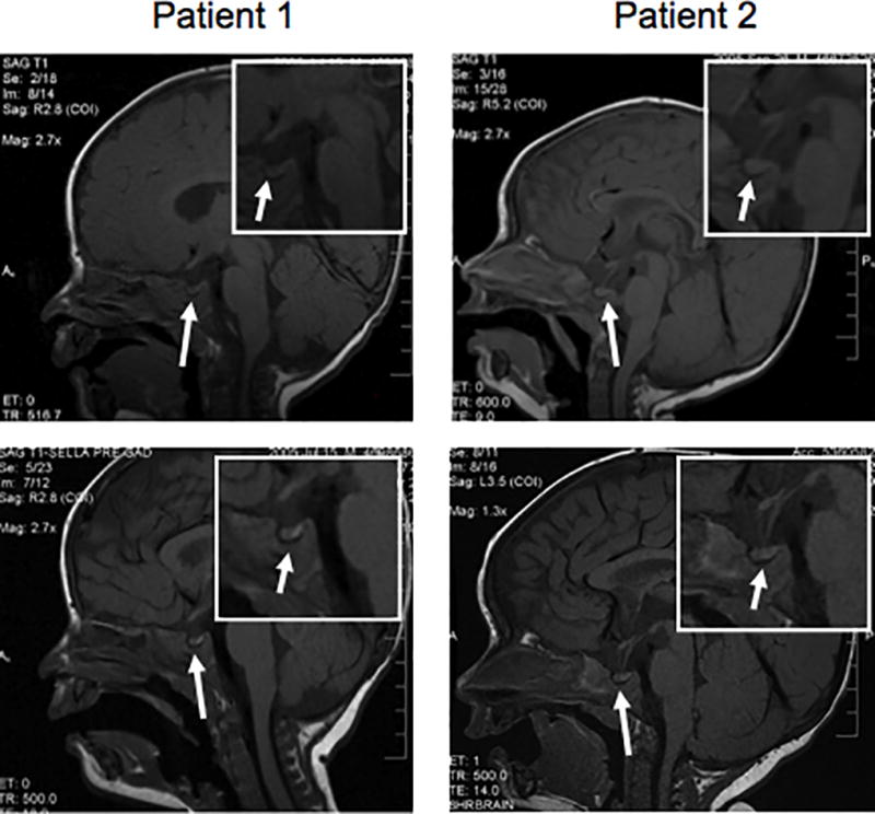
Patients: Two male infants presented with poor growth and hypernatraemia. Both patients had measurable pretreatment serum AVP and polyuria that did not respond to DDAVP, suggesting NDI. However, both also had absent posterior pituitary bright spot on MRI, which is a finding more typical of CDI.
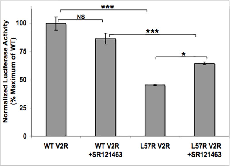
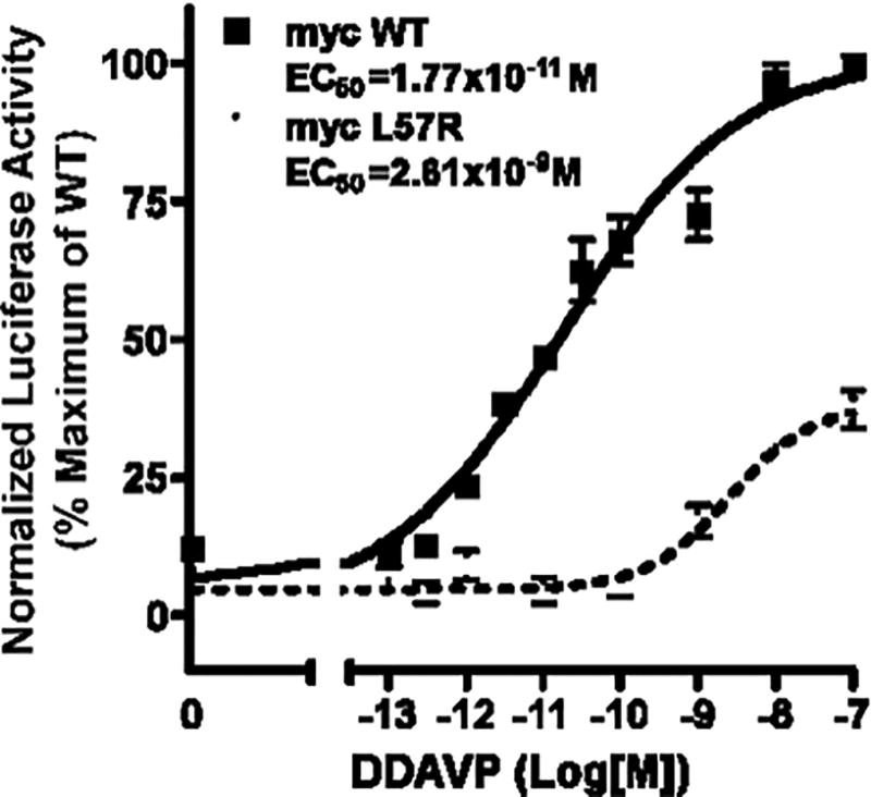
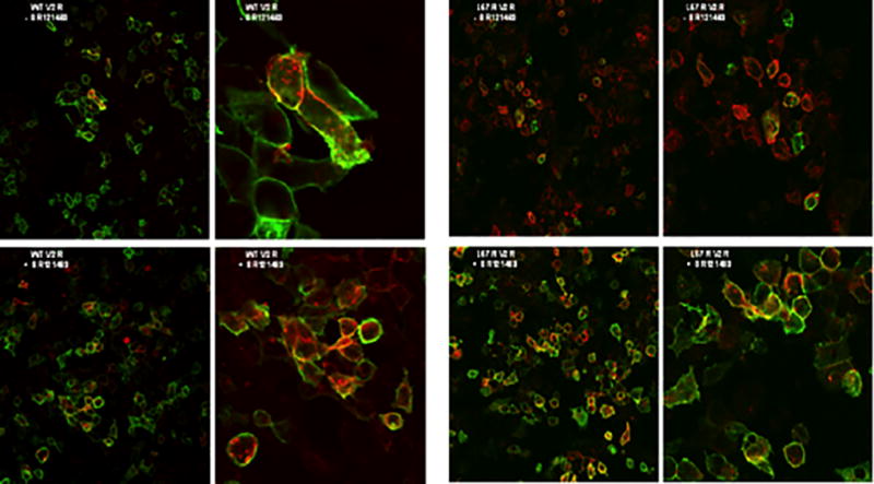
Methods: The AVPR2 gene encoding V2R was sequenced. The identified novel missense mutation was re-created by site-directed mutagenesis and expressed in HEK293 cells. V2R activity was assessed by the ability of transfected cells to produce cAMP in response to stimulation with DDAVP. Membrane localization of V2R was assessed by fluorescence microscopy.
Results: Patient 1 had a deletion of AVPR2; patient 2 had the novel mutation L57R. In transiently transfected HEK293 cells, DDAVP induced detectable but severely impaired L57R V2R activity compared to cells expressing wild-type V2R. Fluorescence microscopy showed that myc-tagged wild-type V2R localized to the cell membrane while L57R V2R remained intracellular. A nonpeptide V2R chaperone, SR121463, partially rescued L57R V2R function by allowing it to reach the cell membrane.
Conclusions: L57R V2R has impaired in vitro activity that can be partially improved by treatment with a V2R chaperone. The posterior pituitary hyperintensity may be absent in infants with XNDI.
Figures
Similar articles
-
V2 vasopressin receptor (V2R) mutations in partial nephrogenic diabetes insipidus highlight protean agonism of V2R antagonists.J Biol Chem. 2012 Jan 13;287(3):2099-106. doi: 10.1074/jbc.M111.268797. Epub 2011 Dec 5. J Biol Chem. 2012. PMID: 22144672 Free PMC article.
-
Functional rescue of vasopressin V2 receptor mutants in MDCK cells by pharmacochaperones: relevance to therapy of nephrogenic diabetes insipidus.Am J Physiol Renal Physiol. 2007 Jan;292(1):F253-60. doi: 10.1152/ajprenal.00247.2006. Epub 2006 Aug 22. Am J Physiol Renal Physiol. 2007. PMID: 16926443
-
Identification and characterization of a novel X-linked AVPR2 mutation causing partial nephrogenic diabetes insipidus: a case report and review of the literature.Metabolism. 2012 Jul;61(7):922-30. doi: 10.1016/j.metabol.2012.01.005. Epub 2012 Mar 3. Metabolism. 2012. PMID: 22386940 Review.
-
Characterization of five novel vasopressin V2 receptor mutants causing nephrogenic diabetes insipidus reveals a role of tolvaptan for M272R-V2R mutation.Sci Rep. 2020 Oct 2;10(1):16383. doi: 10.1038/s41598-020-73089-x. Sci Rep. 2020. PMID: 33009446 Free PMC article.
-
Potential of nonpeptide (ant)agonists to rescue vasopressin V2 receptor mutants for the treatment of X-linked nephrogenic diabetes insipidus.J Neuroendocrinol. 2010 May;22(5):393-9. doi: 10.1111/j.1365-2826.2010.01983.x. Epub 2010 Feb 12. J Neuroendocrinol. 2010. PMID: 20163515 Review.
Cited by
-
Altered agonist sensitivity of a mutant v2 receptor suggests a novel therapeutic strategy for nephrogenic diabetes insipidus.Mol Endocrinol. 2014 May;28(5):634-43. doi: 10.1210/me.2013-1424. Epub 2014 Mar 14. Mol Endocrinol. 2014. PMID: 24628417 Free PMC article.
-
Mutations in the AVPR2, AVP-NPII, and AQP2 genes in Turkish patients with diabetes insipidus.Endocrine. 2012 Dec;42(3):664-9. doi: 10.1007/s12020-012-9704-1. Epub 2012 May 29. Endocrine. 2012. PMID: 22644838
-
Oligomerisation of C. elegans olfactory receptors, ODR-10 and STR-112, in yeast.PLoS One. 2014 Sep 25;9(9):e108680. doi: 10.1371/journal.pone.0108680. eCollection 2014. PLoS One. 2014. PMID: 25254556 Free PMC article.
-
X-Linked Recessive form of Nephrogenic Diabetes Insipidus in a 7-Year-Old Boy.Balkan J Med Genet. 2015 Apr 10;17(2):81-5. doi: 10.2478/bjmg-2014-0078. eCollection 2014 Dec. Balkan J Med Genet. 2015. PMID: 25937802 Free PMC article.
-
Pediatric disorders of water balance.Pediatr Clin North Am. 2011 Oct;58(5):1271-80, xi-xii. doi: 10.1016/j.pcl.2011.07.013. Pediatr Clin North Am. 2011. PMID: 21981960 Free PMC article.
References
-
- Verbalis JG. Diabetes insipidus. Reviews in Endocrine & Metabolic Disorders. 2003;4:177–185. - PubMed
-
- Miller WL. Molecular genetics of familial central diabetes insipidus. Journal of Clinical Endocrinology and Metabolism. 1993;77:592–595. - PubMed
-
- Knoers NV, Deen PM. Molecular and cellular defects in nephrogenic diabetes insipidus. Pediatric Nephrology. 2001;16:1146–1152. - PubMed
-
- Spanakis E, Milord E, Gragnoli C. AVPR2 variants and mutations in nephrogenic diabetes insipidus: review and missense mutation significance. Journal of Cellular Physiology. 2008;217:605–617. - PubMed
Publication types
MeSH terms
Substances
Grants and funding
LinkOut - more resources
Full Text Sources
Miscellaneous
