. 2009 Mar;71(6):1477-95.
doi: 10.1111/j.1365-2958.2009.06616.x. Epub 2009 Jan 23.

Threonine phosphorylation prevents promoter DNA binding of the Group B Streptococcus response regulator CovR

Affiliations

Threonine phosphorylation prevents promoter DNA binding of the Group B Streptococcus response regulator CovR

Wan-Jung Lin et al. Mol Microbiol. 2009 Mar.

Abstract

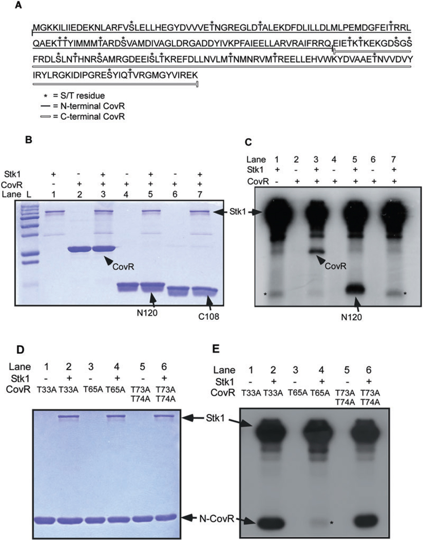
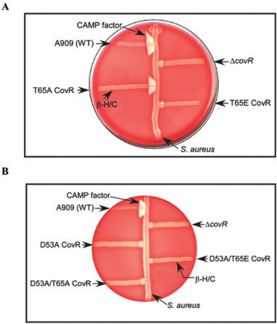
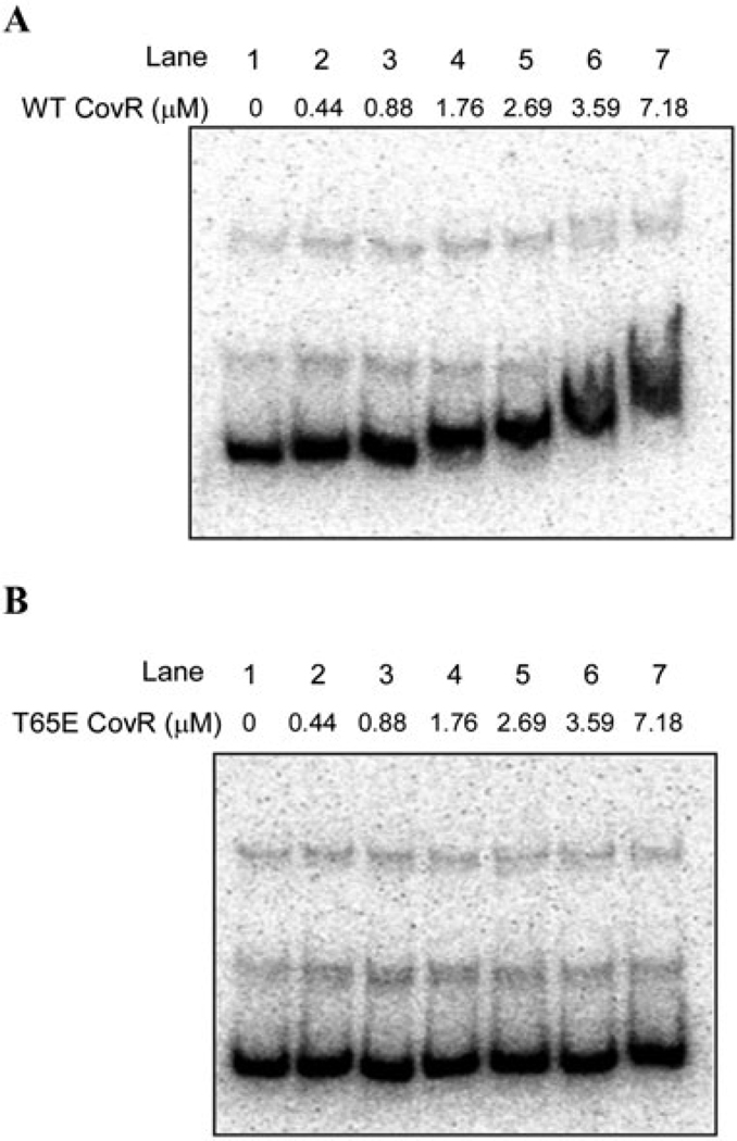
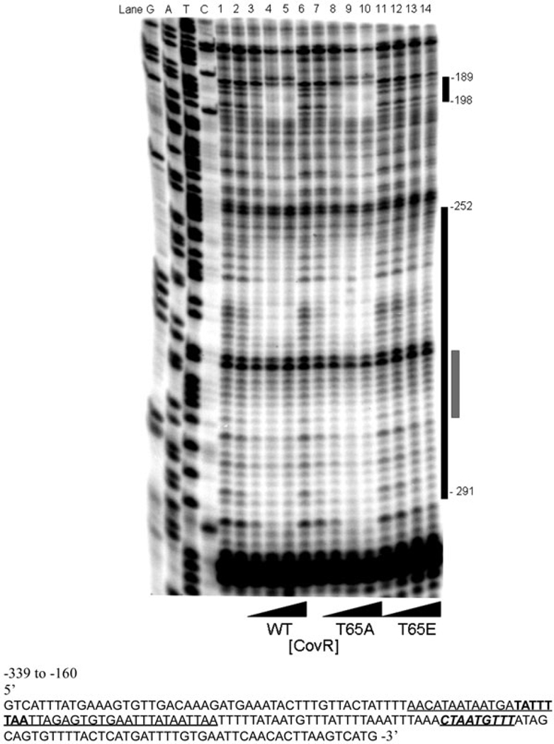
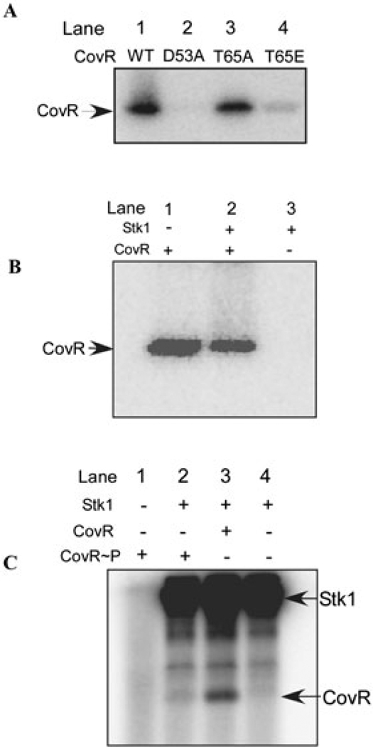
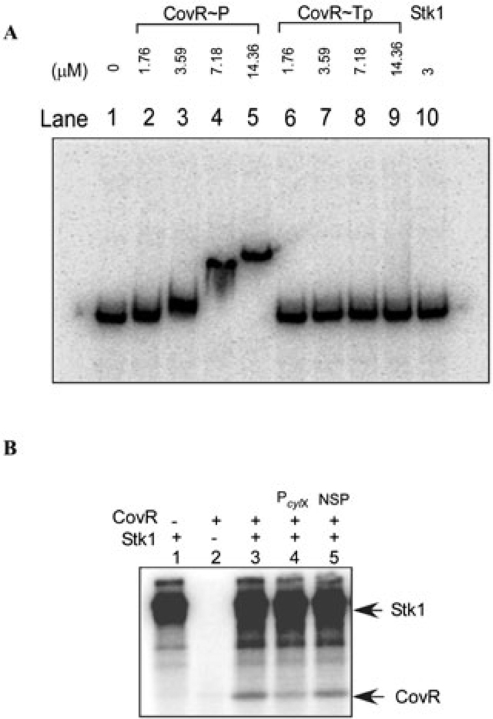
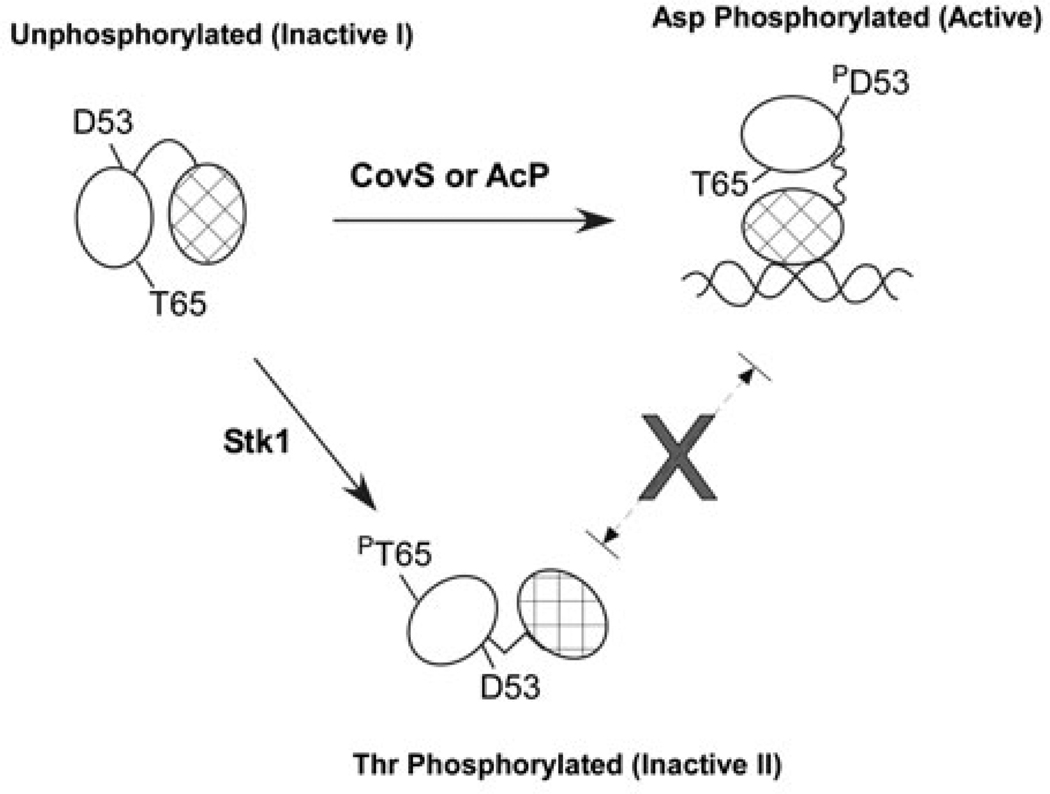
All living organisms communicate with the external environment for their survival and existence. In prokaryotes, communication is achieved by two-component systems (TCS) comprising histidine kinases and response regulators. In eukaryotes, signalling is accomplished by serine/threonine and tyrosine kinases. Although TCS and serine/threonine kinases coexist in prokaryotes, direct cross-talk between these families was first described in Group B Streptococcus (GBS). A serine/threonine kinase (Stk1) and a TCS (CovR/CovS) co-regulate toxin expression in GBS. Typically, promoter binding of regulators like CovR is controlled by phosphorylation of the conserved active site aspartate (D53). In this study, we show that Stk1 phosphorylates CovR at threonine 65. The functional consequence of threonine phosphorylation of CovR in GBS was evaluated using phosphomimetic and silencing substitutions. GBS encoding the phosphomimetic T65E allele are deficient for CovR regulation unlike strains encoding the non-phosphorylated T65A allele. Further, compared with wild-type or T65A CovR, the T65E CovR is unable to bind promoter DNA and is decreased for phosphorylation at D53, similar to Stk1-phosphorylated CovR. Collectively, we provide evidence for a novel mechanism of response regulator control that enables GBS (and possibly other prokaryotes) to fine-tune gene expression for environmental adaptation.


Figures

Fig. 1
Fig. 1
Stk1 phosphorylates CovR at a threonine residue in position 65. A. The entire CovR coding sequence comprising 229 amino acids is shown. The amino acids encoded in the N- (N120) and C-terminal (C108) CovR are indicated by the filled and unfilled line respectively. The N120 CovR contains two serine and six threonine residues, while the C108 CovR (which was constructed to have a methionine start codon) has six serine and eight threonine residues. B and C. In vitro phosphorylation reactions were performed with approximately 0.5–1 µg of GST-Stk1 protein and WT, N120 or C108 CovR as described in the Experimental procedures. All samples were analysed on 15% SDS-PAGE, stained with Coomassie and exposed to autoradiography. Note that Stk1 phosphorylates full-length CovR-His6 and N-terminal CovR-His6 (see lanes 3 and 5 in ‘C’). The band denoted by asterisk (*) observed with C108 (see lane 7 in ‘C’) is also seen with Stk1 alone (see lane 1 in ‘C’). Lane ‘L’ is the pre-stained Protein Standards (Bio-Rad). D and E. In vitro phosphorylation reactions were performed with approximately 0.5–1 µg of GST-Stk1 protein and CovR substitution mutants as described in the Experimental procedures. All samples were analysed on 15% SDS-PAGE, stained with Coomassie and exposed to autoradiography. Autoradiograph shows that Stk1 phosphorylation is drastically reduced or abolished in T65A N-terminal CovR-His6 (Fig. 1E, lane 4) in contrast to T33A (see lane 2 in ‘E’) or T73/74A CovR (see lane 6 in ‘E’). Autophosphorylation of T65A, T33A and T73/74A was not observed (see lanes 1, 3 and 5 in ‘E’).
Fig. 2
Fig. 2
Cytotoxin expression in GBS strains encoding T65A and T65E CovR. A. T65A CovR complements the ΔcovR strain for repression of β-H/C and activation of CAMP factor expression unlike the T65E CovR. CAMP factor activity is represented by the triangular zone of lysis at the junction between GBS and β-lysin producing Staphylococcus aureus on blood-agar plates. β-H/C activity is represented by the zone of clearing around GBS. A909 represents wild-type GBS. The strains encoding the T65A and T65E CovR alleles on the GBS chromosome are isogenic to A909. The ΔcovR is a covR deletion in A909. Note that CAMP factor activity is greater in T65A when compared with T65E or ΔcovR. B. Complementation of the ΔcovR strain by T65A CovR requires D53. GBS strains encoding D53A, D53A/T65A and D53A/T65E CovR are compared with WT and the isogenic ΔcovR strain. Note that the decrease in β-H/C and increase in CAMP factor expression seen in T65A (shown in A) is abolished in D53A/T65A CovR.
Fig. 3
Fig. 3
T65E CovR does not bind promoter DNA. Electrophoretic mobility shift assay was performed using the PcylX promoter DNA as probe. The 176 bp PcylX promoter was amplified using the oligos PcylxL and PcylxR (see Table S1) and incubated with equimolar concentrations of acetyl phosphate treated WT CovR (CovR~P) (A) and T65E CovR~P (B). In both cases, lane 1 represents the probe-only control and lanes 2–7 represent increasing amounts of CovR from 0. 44 to 7.18 µ. Note that retardation in mobility of the probe DNA was observed with increasing concentrations of WT CovR~P and not with the T65E CovR~P.
Fig. 4
Fig. 4
T65E CovR does not protect promoter PcylX from DNase I cleavage. Equimolar concentrations of phosphoramidate-treated WT, T65A and T65E CovR~P were used in DNase I protection assays of the PcylX promoter. The lanes G, A, T and C represent the DNA sequencing ladder. Lanes 1 and 6 represent DNase I-only controls. Lanes 2–5, 7–10 and 11–14 represent WT, T65A and T65E CovR~P at 1, 2, 4 and 8 µM respectively. The co-ordinates of CovR protection from −291 to −252 of the predicted cylX translational start site was described previously (Lamy et al., 2004). These footprinting analyses also indicate CovR protection of PcylX from −198 to −189 which was not previously observed. Importantly, in all cases, the T65A CovR~P demonstrated DNA protection similar to WT CovR, whereas no significant DNA protection is observed with T65E CovR. The black bar to the right of the panel delineates the extent of CovR-mediated protection from DNase I cleavage. The grey bar depicts the predicted CovR DNA sequence motif (Lamy et al., 2004).
Fig. 5
Fig. 5
A. T65E CovR decreases phosphorylation of D53 by [32P]-acetyl phosphate. Equal concentrations of WT, T65A, T65E and D53A CovR were incubated with 32 mM [32P]-acetyl phosphate (specific activity 8.5 mCi mmol−1) at 37°C for 90 min. The samples were subsequently analysed on a 12% SDS-PAGE followed by autoradiography. Note that [32P]-acetyl phosphate-mediated phosphorylation of T65E CovR is significantly reduced (≥ 3-fold) compared with T65A and WT CovR. The experiment was repeated at least three times. B. Stk1 phosphorylation of CovR decreases D53 phosphorylation by [32P]-acetyl phosphate. In vitro phosphorylation of CovR was performed with non-radioactive ATP in the presence and absence of Stk1 prior to incubation with 32 mM [32P]-acetyl phosphate (specific activity 8.5 mCi mmol−1) at 37°C for 90 min. Controls included a reaction containing only Stk1 (lane 3) and no phosphorylation by acetyl phosphate was observed. The samples were subsequently analysed on a 12% SDS-PAGE followed by autoradiography. The figure shown represents one of three independent experiments. Note that [32P]-acetyl phosphate-mediated phosphorylation of CovR is significantly reduced (≥ 2-fold) when phosphorylated by Stk1 (compare lane 2 with lane 1). C. CovR phosphorylation at D53 reduces phosphorylation by Stk1. Equal concentrations of CovR were pre-incubated in the presence and absence of 32 mM acetyl phosphate prior to the addition of Stk1 that was phosphorylated with radioactive ATP (see Experimental procedures). All samples were analysed on 12% SDS-PAGE and exposed to autoradiography. The figure shown represents one of three independent experiments. Lane 4 contains only Stk1 and lane 1 contains only CovR~P. CovR phosphorylation by Stk1 was reduced ≥ 4-fold when pretreated with acetyl phosphate (lane 2) compared with the absence of acetyl phosphate (lane 3).
Fig. 6
Fig. 6
A. Stk1 phosphorylation prevents promoter binding of CovR. In vitro phosphorylation of CovR was performed using Stk1 that was bound to GST-sepharose. Electrophoretic mobility shift assay was then performed using PcylX DNA as probe. The 176 bp PcylX promoter was amplified using the oligos PcylxL and PcylxR (see Table S1) and incubated with equimolar concentrations of threonine-phosphorylated CovR (CovR~Tp) and non-threonine-phosphorylated CovR (see Experimental procedures). Lane 1 represents the probe-only control and lanes 2–5 represent increasing amounts of CovR~P from 1.76 to 14.36 µM and lanes 6–9 represent increasing amounts of CovR~Tp from 1.76 to 14.36 µM. Note that retardation in mobility of the probe DNA was observed with increasing concentrations of CovR~P and not with the CovR~Tp. As an additional control, lane 10 contained 3 µM of Stk1 alone (i.e. no CovR) and demonstrates that Stk1 itself does not bind to PcylX DNA. B. CovR phosphorylation by Stk1 is reduced in the presence of CovR-specific promoter DNA. Equal concentrations of unphosphorylated CovR were pre-incubated with either a CovR-specific promoter, i.e. PcylX (lane 4) or non-specific DNA (lane 5) prior to in vitro phosphorylation reaction with Stk1 (see Experimental procedures). All samples were analysed on 12% SDS-PAGE and exposed to autoradiography. The figure shown is one of three independent experiments. In lane 3, a control reaction that did not contain DNA is shown. CovR phosphorylation was reduced to 60% in the presence of PcylX DNA (lane 4) compared with the absence of DNA (lane 3).
Fig. 7
Fig. 7
Proposed model of Stk1 and CovS regulation of CovR. CovR requires D53 in order to bind DNA and repress or activate transcription of β-H/C and CAMP factor respectively. Phosphorylation by small molecule phosphodonors like acetyl phosphate (AcP) or its cognate kinase CovS converts CovR to the active form. Stk1 phosphorylates the inactive form of CovR, as phosphorylation at D53 or DNA binding decrease the reactivity of T65 to Stk1. As a consequence of T65 phosphorylation, CovR is prevented from binding to DNA and D53 exhibits reduced reactivity.

Similar articles

Cited by

References

    1. Aitken A. Protein consensus sequence motifs. Methods Mol Biol. 2003;211:465–485. - PubMed
    1. Ames SK, Frankema N, Kenney LJ. C-terminal DNA binding stimulates N-terminal phosphorylation of the outer membrane protein regulator OmpR from Escherichia coli. Proc Natl Acad Sci USA. 1999;96:11792–11797. - PMC - PubMed
    1. Ausubel FM, Brent R, Kingston RE, Moore DD, Seidman JG, Smith JA, Struhl K. Current Protocols in Molecular Biology. New York: John Wiley & Sons; 1997.
    1. Baker CJ, Edwards MW. Group B streptococcal infections. In: Remington JS, Klein JO, editors. Infectious Diseases of the Fetus and Newborn Infant. Philadelphia, PA: W.B. Saunders; 1995. pp. 980–1054.
    1. Barz C, Abahji TN, Trulzsch K, Heesemann J. The Yersinia Ser/Thr protein kinase YpkA/YopO directly interacts with the small GTPases RhoA and Rac-1. FEBS Lett. 2000;482:139–143. - PubMed

Publication types

MeSH terms