Review
. 2009 Jan 26:7:8.
doi: 10.1186/1477-7827-7-8.

New discoveries on the biology and detection of human chorionic gonadotropin

Affiliations
Review

New discoveries on the biology and detection of human chorionic gonadotropin

Laurence A Cole. Reprod Biol Endocrinol. .

Abstract

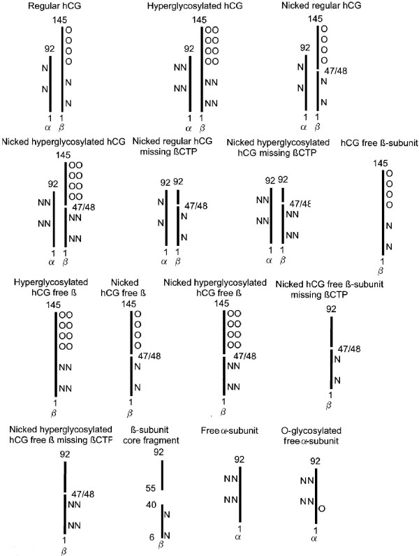
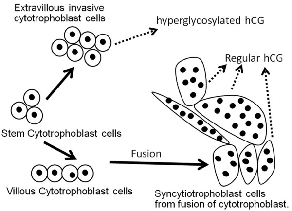
Human chorionic gonadotropin (hCG) is a glycoprotein hormone comprising 2 subunits, alpha and beta joined non covalently. While similar in structure to luteinizing hormone (LH), hCG exists in multiple hormonal and non-endocrine agents, rather than as a single molecule like LH and the other glycoprotein hormones. These are regular hCG, hyperglycosylated hCG and the free beta-subunit of hyperglycosylated hCG. For 88 years regular hCG has been known as a promoter of corpus luteal progesterone production, even though this function only explains 3 weeks of a full gestations production of regular hCG. Research in recent years has explained the full gestational production by demonstration of critical functions in trophoblast differentiation and in fetal nutrition through myometrial spiral artery angiogenesis. While regular hCG is made by fused villous syncytiotrophoblast cells, extravillous invasive cytotrophoblast cells make the variant hyperglycosylated hCG. This variant is an autocrine factor, acting on extravillous invasive cytotrophoblast cells to initiate and control invasion as occurs at implantation of pregnancy and the establishment of hemochorial placentation, and malignancy as occurs in invasive hydatidiform mole and choriocarcinoma. Hyperglycosylated hCG inhibits apoptosis in extravillous invasive cytotrophoblast cells promoting cell invasion, growth and malignancy. Other non-trophoblastic malignancies retro-differentiate and produce a hyperglycosylated free beta-subunit of hCG (hCG free beta). This has been shown to be an autocrine factor antagonizing apoptosis furthering cancer cell growth and malignancy. New applications have been demonstrated for total hCG measurements and detection of the 3 hCG variants in pregnancy detection, monitoring pregnancy outcome, determining risk for Down syndrome fetus, predicting preeclampsia, detecting pituitary hCG, detecting and managing gestational trophoblastic diseases, diagnosing quiescent gestational trophoblastic disease, diagnosing placental site trophoblastic tumor, managing testicular germ cell malignancies, and monitoring other human malignancies. There are very few molecules with such wide and varying functions as regular hCG and its variants, and very few tests with such a wide spectrum of clinical applications as total hCG.


Figures

Figure 1
Figure 1
Amino acid sequence of hCG α-subunit and β-subunit [16,17]. Digits indicated amino acid residue positions and N and O indicate the positions of N- and O-linked oligosaccharides.
Figure 2
Figure 2
Structures of O-linked hexa- and tris-saccharide and N-linked Bi (biantennary) and Tri (triantennary) oligosaccharides attached to regular hCG, hyperglycosylated hCG and hyperglycosylated hCG free β [18-20]. NeuAc is N-acetylneuraminic acid or sialic acid, GalNAc in N-acetylgalactosamine, Gal is galactose, GlcNAc is N-acetylglucosamine, Man is mannose and Fuc is fucose.
Figure 3
Figure 3
Outline of the structures of the 15 common hCG variants present in serum and urine samples in either pregnancy, gestational trophoblastic disease or other malignancy. Numbers refer to subunit polypeptide amino acid numbers (as in 1 and 145 in the 145 amino acid long β-subunit), O refers to O-linked and N to N-linked oligosaccharides. OO and NN refer to large or hyperglycosylated oligosaccharides. α is α-subunit and β is β-subunit. βCTP is the C-terminal segment (residues 93–145) on the regular or hyperglycosylated hCG β-subunit.
Figure 4
Figure 4
The differentiation of trophoblast cells in placental villi [54]. Fusion of villous cytotrophoblast cells is controlled by regular hCG [55]. Extravillous invasive cytotrophoblast cells produce hyperglycosylated hCG [44], while fused syncytiotrophoblast cells make regular hCG [40-42].
Figure 5
Figure 5
The dissociation, degradation and clearance pathways of regular hCG [39,78-87]. Metabolic clearance half-times (MCR) are those published by Wehman and colleagues [85-88]. Similar degradation and clearance pathways are predicted for hyperglycosylated hCG and hyperglycosylated hCG free β with an end product of urine β-subunit core fragment.
Figure 6
Figure 6
Villous placental tissue, cytotrophoblast and syncytiotrophoblast cells, and regular hCG and hyperglycosylated hCG function. Panel A illustrated blastocyst implantation and engulfment and trophoblast invasion at 3–5 weeks of gestation. Arrows illustrates the biological functions of regular hCG and hyperglycosylated hCG. Mononuclear cells are cytotrophoblast cells, cells with multiple nuclei (black circles) represen syncytiotrophoblast cells. In Panel B villous trophoblast formation and function are illustrated at 6–8 weeks of gestation [44,54,114,115]. Figure illustrates villous trophoblast (anchoring villus and floating villus) growth 5 to 10 weeks of gestation, and invasion of the decidua and myometrium in establishing hemochorial placentation. Panel C illustrates functional hemochorial placentation in floating villus at 10–12 weeks of gestation after invasion is complete [114-118]. Cells with varying numbers of multiple nuclei (black circles) represent villous syncytiotrophoblast (VSyn), mononuclear cells are villous cytotrophoblast (VCyto) and extravillous invasive cytotrophoblast (EVICyto). Arrows shows biological actions of regular hCG and hyperglycosylated hCG.

References

    1. Hirose T. Exogenous stimulation of corpus luteum formation in the rabbit: influence of extracts of human placenta, decidua, fetus, hydatid mole, and corpus luteum on the rabbit gonad. J Jpn Gynecol Soc. 1920;16:1055.
    1. Zondek B, Aschheim S. The Zondek-Ascheim Pregnancy Test. Can Med Assoc J. 1930;22:251–253. - PMC - PubMed
    1. Aschheim S, Zondek B. Das Hormon des hypophysenvorderlappens: testobjekt zum Nachweis des hormons. Klin Wochenschr. 1927;6:248–252.
    1. Wide L, Gemzell CA. An immunological pregnancy test. Acta Endocrinol. 1960;35:261–267. - PubMed
    1. Rushworth AG, Orr AH, Bagshawe KD. The concentration of HCG in the plasma and spinal fluid of patients with trophoblastic tumours in the central nervous system. Br J Cancer. 1968;22:253–257. - PMC - PubMed

MeSH terms

Substances