

Upregulation of prolylcarboxypeptidase (PRCP) in lipopolysaccharide (LPS) treated endothelium promotes inflammation
- PMID: 19171072
- PMCID: PMC2639534
- DOI: 10.1186/1476-9255-6-3
Upregulation of prolylcarboxypeptidase (PRCP) in lipopolysaccharide (LPS) treated endothelium promotes inflammation
Abstract
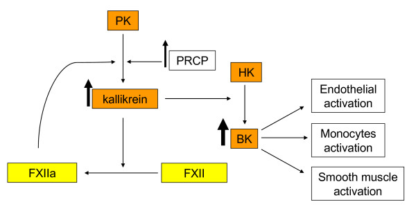
Background: Prolylcarboxypeptidase (Prcp) gene, along with altered PRCP and kallikrein levels, have been implicated in inflammation pathogenesis. PRCP regulates angiotensin 1-7 (Ang 1-7) - and bradykinin (BK) - stimulated nitric oxide production in endothelial cells. The mechanism through which kallikrein expression is altered during infection is not fully understood. Investigations were performed to determine the association between PRCP and kallikrein levels as a function of the upregulation of PRCP expression and the link between PRCP and inflammation risk in lipopolysaccharide (LPS)-induced endothelium activation.
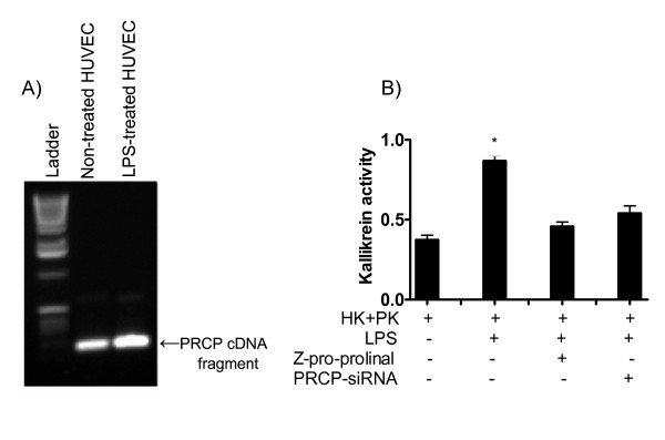
Methods: The Prcp transcript expression in LPS-induced human umbilical vein endothelial cells (HUVEC) activation was determined by RT-PCR for mRNA. PRCP-dependent kallikrein pathway was determined either by Enzyme Linked ImmunoSorbent Assay (ELISA) or by biochemical assay.
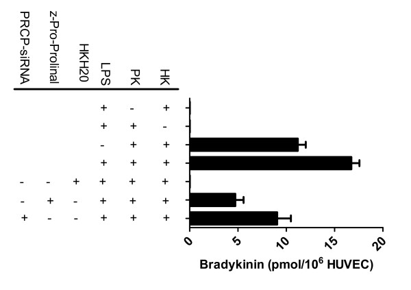
Results: We report that PRCP is critical to the maintenance of the endothelial cells, and its upregulation contributes to the risk of developing inflammation. Significant elevation in kallikrein was seen on LPS-treated HUVECs. The conversion of PK to kallikrein was blocked by the inhibitor of PRCP, suggesting that PRCP might be a risk factor for inflammation.
Conclusion: The increased PRCP lead to a sustained production of bradykinin in endothelium following LPS treatment. This amplification may be an additional mechanism whereby PRCP promotes a sustained inflammatory response. A better appreciation of the role of PRCP in endothelium may contribute to a better understanding of inflammatory vascular disorders and to the development of a novel treatment.
Figures
Similar articles
-
Increased Prolylcarboxypeptidase Expression Can Serve as a Biomarker of Senescence in Culture.Molecules. 2024 May 9;29(10):2219. doi: 10.3390/molecules29102219. Molecules. 2024. PMID: 38792081 Free PMC article.
-
Identification and characterization of prolylcarboxypeptidase as an endothelial cell prekallikrein activator.J Biol Chem. 2002 May 17;277(20):17962-9. doi: 10.1074/jbc.M106101200. Epub 2002 Feb 5. J Biol Chem. 2002. PMID: 11830581
-
Recombinant prolylcarboxypeptidase activates plasma prekallikrein.Blood. 2004 Jun 15;103(12):4554-61. doi: 10.1182/blood-2003-07-2510. Epub 2004 Mar 2. Blood. 2004. PMID: 14996700
-
Prolylcarboxypeptidase: a cardioprotective enzyme.Int J Biochem Cell Biol. 2009 Mar;41(3):477-81. doi: 10.1016/j.biocel.2008.02.022. Epub 2008 Feb 29. Int J Biochem Cell Biol. 2009. PMID: 18396440 Review.
-
Prolylcarboxypeptidase (PrCP) inhibitors and the therapeutic uses thereof: a patent review.Expert Opin Ther Pat. 2017 Oct;27(10):1077-1088. doi: 10.1080/13543776.2017.1349104. Epub 2017 Jul 12. Expert Opin Ther Pat. 2017. PMID: 28699813 Review.
Cited by
-
Prolyl Carboxypeptidase Mediates the C-Terminal Cleavage of (Pyr)-Apelin-13 in Human Umbilical Vein and Aortic Endothelial Cells.Int J Mol Sci. 2021 Jun 22;22(13):6698. doi: 10.3390/ijms22136698. Int J Mol Sci. 2021. PMID: 34206648 Free PMC article.
-
Increased Expression of NOTCH-1 and T Helper Cell Transcription Factors in Patients with Acquired Aplastic Anemia.Iran Biomed J. 2023 Nov 1;27(6):357-65. doi: 10.61186/ibj.3754. Epub 2022 Jul 31. Iran Biomed J. 2023. PMID: 37980558 Free PMC article.
-
Prolyl carboxypeptidase activity in the circulation and its correlation with body weight and adipose tissue in lean and obese subjects.PLoS One. 2018 May 17;13(5):e0197603. doi: 10.1371/journal.pone.0197603. eCollection 2018. PLoS One. 2018. PMID: 29772029 Free PMC article.
-
Pro-opiomelanocortin and its Processing Enzymes Associate with Plaque Stability in Human Atherosclerosis - Tampere Vascular Study.Sci Rep. 2018 Oct 10;8(1):15078. doi: 10.1038/s41598-018-33523-7. Sci Rep. 2018. PMID: 30305673 Free PMC article.
-
Bradykinin B1 receptor signaling triggers complement activation on endothelial cells.Front Immunol. 2025 Feb 7;16:1527065. doi: 10.3389/fimmu.2025.1527065. eCollection 2025. Front Immunol. 2025. PMID: 39991158 Free PMC article.
References
-
- Tamaoki J, Sugimoto F, Tagaya E, Isono K, Chiyotani A, Konno K. Angiotensin II 1 receptor-mediated contraction of pulmonary artery and its modulation by prolylcarboxypeptidase. J Appl Physiol. 1994;76:1439–1444. - PubMed
-
- Mallela J, Yang J, Shariat-Madar Z. Prolylcarboxypeptidase: A cardioprotective enzyme. Int J Biochem Cell Biol. 2008 - PubMed
-
- Zhao Y, Qiu Q, Mahdi F, Shariat-Madar Z, Rojkjaer R, Schmaier AH. Assembly and activation of HK-PK complex on endothelial cells results in bradykinin liberation and NO formation. Am J Physiol Heart Circ Physiol. 2001;280:H1821–H1829. - PubMed
-
- Colman RW, Schmaier AH. Contact system: a vascular biology modulator with anticoagulant, profibrinolytic, antiadhesive, and proinflammatory attributes. Blood. 1997;90:3819–3843. - PubMed
Grants and funding
LinkOut - more resources
Full Text Sources
Miscellaneous
