Truncated dystrophins can influence neuromuscular synapse structure
- PMID: 19171194
- PMCID: PMC2826111
- DOI: 10.1016/j.mcn.2008.12.011
Truncated dystrophins can influence neuromuscular synapse structure
Abstract
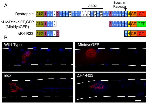
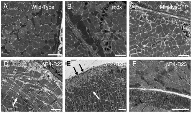
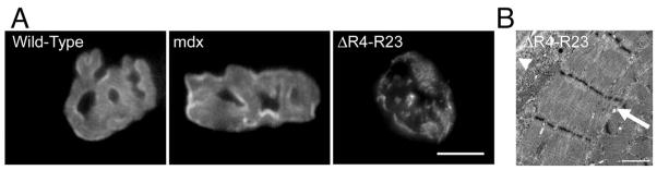
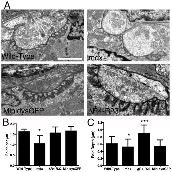
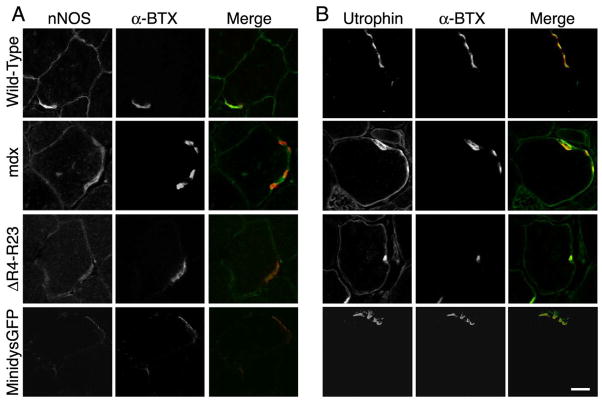
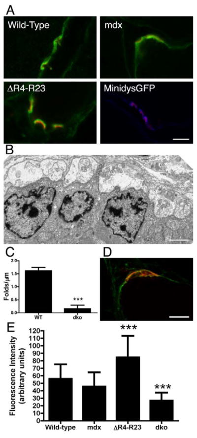
Duchenne muscular dystrophy (DMD) is characterized by muscle degeneration and structural defects in the neuromuscular synapse that are caused by mutations in dystrophin. Whether aberrant neuromuscular synapse structure is an indirect consequence of muscle degeneration or a direct result of loss of dystrophin function is not known. Rational design of truncated dystrophins has enabled the design of expression cassettes highly effective at preventing muscle degeneration in mouse models of DMD using gene therapy. Here we examined the functional capacity of a minidystrophin (minidysGFP) and a microdystrophin (microdystrophin(DeltaR4-R23)) transgene on the maturation and maintenance of neuromuscular junctions (NMJ) in mdx mice. We found that minidysGFP prevents fragmentation and the loss of postsynaptic folds at the NMJ. In contrast, microdystrophin (DeltaR4-R23) was unable to prevent synapse fragmentation in the limb muscles despite preventing muscle degeneration, although fragmentation was observed to temporally correlate with the formation of ringed fibers. Surprisingly, microdystrophin(DeltaR4-R23) increased the length of synaptic folds in the diaphragm muscles of mdx mice independent of muscle degeneration or the formation of ringed fibers. We also demonstrate that the number and depth of synaptic folds influences the density of voltage-gated sodium channels at the neuromuscular synapse in mdx, microdystrophin(DeltaR4-R23)/mdx and mdx:utrophin double knockout mice. Together, these data suggest that maintenance of the neuromuscular synapse is governed through its lateral association with the muscle cytoskeleton, and that dystrophin has a direct role in promoting the maturation of synaptic folds to allow more sodium channels into the junction.
Figures
Similar articles
-
C-terminal-truncated microdystrophin recruits dystrobrevin and syntrophin to the dystrophin-associated glycoprotein complex and reduces muscular dystrophy in symptomatic utrophin/dystrophin double-knockout mice.Mol Ther. 2006 Jul;14(1):79-87. doi: 10.1016/j.ymthe.2006.01.007. Epub 2006 Mar 23. Mol Ther. 2006. PMID: 16563874 Free PMC article.
-
Characterization of neuromuscular synapse function abnormalities in multiple Duchenne muscular dystrophy mouse models.Eur J Neurosci. 2016 Jun;43(12):1623-35. doi: 10.1111/ejn.13249. Epub 2016 May 9. Eur J Neurosci. 2016. PMID: 27037492
-
Delivery of AAV2/9-microdystrophin genes incorporating helix 1 of the coiled-coil motif in the C-terminal domain of dystrophin improves muscle pathology and restores the level of α1-syntrophin and α-dystrobrevin in skeletal muscles of mdx mice.Hum Gene Ther. 2011 Nov;22(11):1379-88. doi: 10.1089/hum.2011.020. Epub 2011 May 25. Hum Gene Ther. 2011. PMID: 21453126 Free PMC article.
-
Alterations of neuromuscular junctions in Duchenne muscular dystrophy.Neurosci Lett. 2020 Oct 15;737:135304. doi: 10.1016/j.neulet.2020.135304. Epub 2020 Aug 17. Neurosci Lett. 2020. PMID: 32818587 Free PMC article. Review.
-
Dystrophin and utrophin: genetic analyses of their role in skeletal muscle.Microsc Res Tech. 2000 Feb 1-15;48(3-4):155-66. doi: 10.1002/(SICI)1097-0029(20000201/15)48:3/4<155::AID-JEMT4>3.0.CO;2-0. Microsc Res Tech. 2000. PMID: 10679963 Review.
Cited by
-
Uncovering molecular biomarkers that correlate cognitive decline with the changes of hippocampus' gene expression profiles in Alzheimer's disease.PLoS One. 2010 Apr 13;5(4):e10153. doi: 10.1371/journal.pone.0010153. PLoS One. 2010. PMID: 20405009 Free PMC article.
-
Nanotherapy for Duchenne muscular dystrophy.Wiley Interdiscip Rev Nanomed Nanobiotechnol. 2018 Mar;10(2):10.1002/wnan.1472. doi: 10.1002/wnan.1472. Epub 2017 Apr 11. Wiley Interdiscip Rev Nanomed Nanobiotechnol. 2018. PMID: 28398005 Free PMC article. Review.
-
Microutrophin expression in dystrophic mice displays myofiber type differences in therapeutic effects.PLoS Genet. 2020 Nov 11;16(11):e1009179. doi: 10.1371/journal.pgen.1009179. eCollection 2020 Nov. PLoS Genet. 2020. PMID: 33175853 Free PMC article.
-
Evaluation of the dystrophin carboxy-terminal domain for micro-dystrophin gene therapy in cardiac and skeletal muscles in the DMDmdx rat model.Gene Ther. 2022 Sep;29(9):520-535. doi: 10.1038/s41434-022-00317-6. Epub 2022 Feb 1. Gene Ther. 2022. PMID: 35105949
-
The polyproline site in hinge 2 influences the functional capacity of truncated dystrophins.PLoS Genet. 2010 May 20;6(5):e1000958. doi: 10.1371/journal.pgen.1000958. PLoS Genet. 2010. PMID: 20502633 Free PMC article.
References
-
- Abmayr S, Chamberlain JS. The structure and function of dystrophin. In: Winder S, editor. The Molecular Mechanisms of Muscular Dystrophy. Landes Bioscience; Georgetown: 2006.
-
- Anderson JL, Head SI, Rae C, Morley JW. Brain function in Duchenne muscular dystrophy. Brain. 2002;125:4–13. - PubMed
Publication types
MeSH terms
Substances
Grants and funding
LinkOut - more resources
Full Text Sources
Medical
Molecular Biology Databases
