Review
. 2009 May 15;587(Pt 10):2163-77.
doi: 10.1113/jphysiol.2008.165860. Epub 2009 Jan 26.

Phenomics of cardiac chloride channels: the systematic study of chloride channel function in the heart

Affiliations
Review

Phenomics of cardiac chloride channels: the systematic study of chloride channel function in the heart

Dayue Duan. J Physiol. .

Abstract

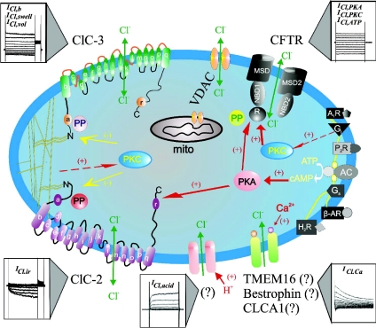
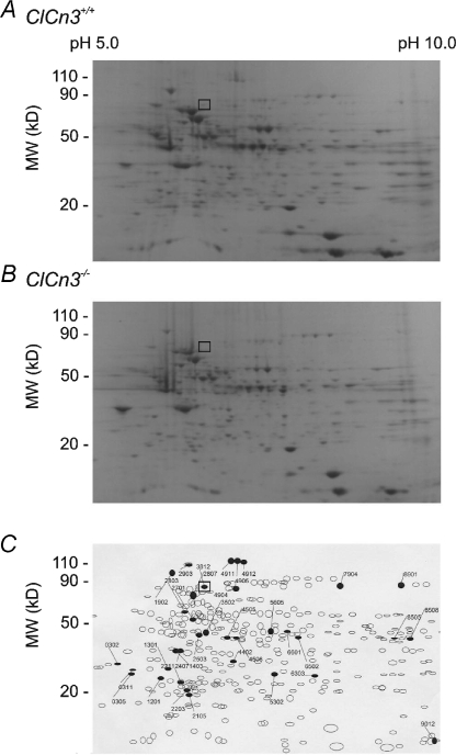
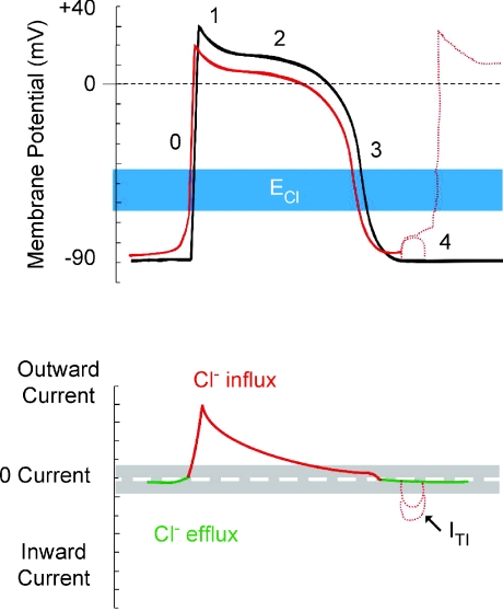
Recent studies have identified several chloride (Cl-) channel genes in the heart, including CFTR, ClC-2, ClC-3, CLCA, Bestrophin, and TMEM16A. Gene targeting and transgenic techniques have been used to delineate the functional role of cardiac Cl- channels in the context of health and disease. It has been shown that Cl- channels may contribute to cardiac arrhythmogenesis, myocardial hypertrophy and heart failure, and cardioprotection against ischaemia-reperfusion. The study of physiological or pathophysiological phenotypes of cardiac Cl- channels, however, may be complicated by the compensatory changes in the animals in response to the targeted genetic manipulation. Alternatively, tissue-specific conditional or inducible knockout or knockin animal models may be more valuable in the phenotypic studies of specific Cl- channels by limiting the effect of compensation on the phenotype. The integrated function of Cl- channels may involve multi-protein complexes of the Cl- channel subproteome and similar phenotypes can be attained from alternative protein pathways within cellular networks, which are influenced by genetic and environmental factors. Therefore, the phenomics approach, which characterizes phenotypes as a whole phenome and systematically studies the molecular changes that give rise to particular phenotypes achieved by modifying the genotype (such as gene knockouts or knockins) under the scope of genome/proteome/phenome, may provide a more complete understanding of the integrated function of each cardiac Cl- channel in the context of health and disease.


Figures

Figure 1
Figure 1. Schematic representation of Cl channels in cardiac myocytes
Cl channels and their corresponding molecular entities or candidates are indicated. ClC-3, a member of voltage-gated ClC Cl channel family, encodes Cl channels that are volume regulated (ICl,vol) and can be activated by cell swelling (ICl,swell) induced by exposure to hypotonic extracellular solutions or possibly membrane stretch. ICl,b is a basally activated ClC-3 Cl current. ClC-2, a member of voltage-gated ClC Cl channel family, is responsible for a volume-regulated and hyperpolarization-activated inward rectifying Cl current (ICl,ir). Membrane topology models (α-helices a-r) for ClC-3 and ClC-2 are modified from Dutzler et al. (2002). ICl,acid is a Cl current regulated by extracellular pH and the molecular entity for ICl,acid is currently unknown. ICl,Ca is a Cl current activated by increased intracellular Ca2+ concentration ([Ca2+]i); Molecular candidates for ICl,Ca include CLCA1, a member of a Ca2+-sensitive Cl channel family (CLCA), bestrophin-2, a member of the bestrophin gene family, and TMEM16, transmembrane protein 16. CFTR, cystic fibrosis transmembrane conductance regulator, encodes Cl channels activated by stimulation of cAMP–protein kinase A (PKA) pathway (ICl,PKA), protein kinase C (PKC) (ICl,PKC), or extracellular ATP through purinergic receptors (ICl,ATP). CFTR is composed of two membrane spanning domains (MSD1 and MSD2), two nucleotide binding domains (NBD1 and NBD2) and a regulatory subunit (R). P, phosphorylation sites for PKA and PKC; PP, serine–threonine protein phosphatases; Gi, heterodimeric inhibitory G protein; A1R, adenosine type 1 receptor; AC, adenylyl cyclase; H2R, histamine type II receptor; Gs, heterodimeric stimulatory G protein; β-AR, β-adrenergic receptor; P2R, purinergic type 2 receptor; proposed intracellular signalling pathway for purinergic activation of CFTR. VDAC, voltage-dependent anion channels (porin); mito, mitochondrion.
Figure 3
Figure 3. Modulation of cardiac electrical activity by activation of ClC-2 channels in cardiac pacemaker cells and myocytes
Changes in action potentials (top panels) and membrane currents (bottom panels) of cardiac pacemaker cells (A), or atrial and ventricular myocytes (B), due to activation of ClC-2 channels are depicted. ICl.ir is activated by hyperpolarization, cell swelling, and acidosis. Top panels, numbers illustrate conventional phases of a prototype ventricular action potential under control conditions (black) and after activation of ICl (red). Range of estimates for normal physiological values for Cl equilibrium potential (ECl) is indicated in blue. Bottom panels, range of zero-current values corresponding to ECl is shown in grey. A, activation of ICl.ir in pacemaker cells during hyperpolarization causes acceleration of phase 4 depolarization and automaticity, shortening of action potential duration, and decrease in cycle length and action potential amplitude (dashed red line in top panel). B, activation of ICl.ir in atrial and ventricular myocytes during hyperpolarization causes depolarization of resting membrane potential and induction of phase 4 autodepolarization and abnormal electrical impulse (trigger activity) and automaticity (dotted red line in top panel).
Figure 2
Figure 2. Modulation of cardiac electrical activity by activation of Cl channels in heart
Changes in action potentials (top), membrane currents (middle), and ECG (bottom) due to activation of CFTR or volume-regulated ClC-3 Cl channels are depicted. Top panel, numbers illustrate conventional phases of a prototype ventricular action potential under control conditions (black) and after activation of ICl (red). Range of estimates for normal physiological values for Cl equilibrium potential (ECl) is indicated in blue. Middle panel, range of zero-current values corresponding to ECl is shown in grey. Activation of CFTR or ClC-3 channels generates both inward (indicated by green) and outward (indicated by red) currents and cause both depolarization as well as repolarization during the action potential. Activation of ICl, therefore, induces larger membrane depolarization and induction of early afterdepolarizations (EAD) under conditions where resting K+ conductance is reduced (dotted red lines in top panel). Bottom panel, the letters (P, Q, R, S and T) indicate the conventional waves of ECG complex under control conditions (black) and after activation of ICl (red). Corresponding to the shortening of action potential in ventricular myocytes activation of ICl causes a shortening of Q–T interval. See text for details.
Figure 4
Figure 4. Comparison of pressure overload-induced remodelling of wild-type and Clcn3−/− mouse hearts
Hearts from age-matched wild-type (WT, Clcn3+/+) and Clcn3−/− mice were excised 1 week (top panel) or 10 weeks (bottom panel) after minimally invasive transverse aorta binding (MTAB) or sham operation are shown. Hearts were cleaned of blood and connective tissues and then fixed in 4% paraformaldehyde. Bar = 5 mm. Compared to WT mice disruption of ClC-3 gene significantly changed the remodelling process after MTAB. Both left ventricle and atrium were extremely enlarged after 10 weeks of MTAB.
Figure 5
Figure 5. Comparative two-dimensional electrophoresis analysis of protein expression patterns in membranes of cardiac cells from Clcn3+/+ and Clcn3−/− mice
A, representative 2-D gel depicts Coomassie-stained proteins from wild-type (Clcn3+/+) mouse heart. B, representative 2-D gel depicts Coomassie-stained proteins from Clcn3−/− mouse heart. C, spot sets created from images of 2-D gels of both wild-type and Clcn3−/− mouse heart run under the same conditions as the gels in A and B and compared using Bio-Rad PDQuest version 7.1.1 software. Three gels were run for each mouse heart type; two hearts were pooled to provide proteins for each gel. The filled symbols indicate changes in protein patterns in Clcn3−/− compared to wild-type. A total of 35 proteins consistently changed (minimum criteria: > 2-fold change) in membranes from Clcn3−/− mouse heart in all 3 experiments (6 missing proteins, 2 new proteins, 9 up-regulated proteins, 15 down-regulated proteins, and 2 translocated proteins). The open squares (□) in A, B and C indicate the location (molecular mass 85 kDa and pI 6.9) of the ClC-3 protein spot (No. 3812) in the 2-D gels, which was independently confirmed by Western blotting using a specific anti-ClC-3 C670−687 antibody. (From Yamamoto-Mizuma et al. 2004b).
Figure 6
Figure 6. Modulation of cardiac electrical activity by activation of Ca2+-activated Cl channels in heart
Changes in action potentials (top) and membrane currents (bottom) due to activation of Ca2+-activated Cl channels are depicted. Top panel, numbers illustrate conventional phases of a prototype ventricular action potential under control conditions (black) and after activation of ICl (red). Range of estimates for normal physiological values for ECl is indicated in blue. Bottom panel, range of zero-current values corresponding to ECl is shown in grey. Activation of ICl.Ca during [Ca2+]i overload results in oscillatory transient inward current (ITI) and induction of delayed afterdepolarization (DAD) (dotted red lines).

Similar articles

Cited by

References

    1. Bahinski A, Nairn AC, Greengard P, Gadsby DC. Chloride conductance regulated by cyclic AMP-dependent protein kinase in cardiac myocytes. Nature. 1989;340:718–721. - PubMed
    1. Baker MA, Lane DJ, Ly JD, De Pinto V, Lawen A. VDAC1 is a transplasma membrane NADH-ferricyanide reductase. J Biol Chem. 2004;279:4811–4819. - PubMed
    1. Batthish M, Diaz RJ, Zeng HP, Backx PH, Wilson GJ. Pharmacological preconditioning in rabbit myocardium is blocked by chloride channel inhibition. Cardiovasc Res. 2002;55:660–671. - PubMed
    1. Baumgarten CM, Clemo HF. Swelling-activated chloride channels in cardiac physiology and pathophysiology. Prog Biophys Mol Biol. 2003;82:25–42. - PubMed
    1. Baumgarten CM, Fozzard HA. Intracellular chloride activity in mammalian ventricular muscle. Am J Physiol Cell Physiol. 1981;241:C121–C129. - PubMed

Publication types

MeSH terms

Substances

LinkOut - more resources