

Pathophysiology and puzzles of the volume-sensitive outwardly rectifying anion channel
- PMID: 19171657
- PMCID: PMC2697288
- DOI: 10.1113/jphysiol.2008.165076
Pathophysiology and puzzles of the volume-sensitive outwardly rectifying anion channel
Abstract
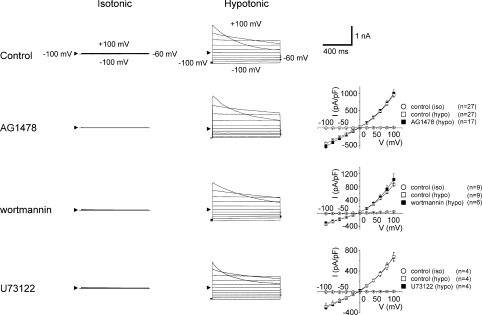
Cell swelling activates or upregulates a number of anion channels. Of the volume-activated or -regulated anion channels (VAACs or VRACs), the volume-sensitive outwardly rectifying anion channel (VSOR) is most prominently activated and ubiquitously expressed. This channel is known to be involved in a variety of physiological processes including cell volume regulation, cell proliferation, differentiation and cell migration as well as cell turnover involving apoptosis. Recent studies have shown that VSOR activity is also involved in a number of pathophysiological processes including the acquisition of cisplatin resistance by cancer cells, ischaemia-reperfusion-induced death of cardiomyocytes and hippocampal neurons, glial necrosis under lactacidosis as well as neuronal necrosis under excitotoxicity. Moreover, VSOR serves as the pathway for glutamate release from astrocytes under ischaemic conditions and when stimulated by bradykinin, an initial mediator of inflammation. So far, many signalling molecules including the EGF receptor, PI3K, Src, PLCgamma and Rho/Rho kinase have been implicated in the regulation of VSOR activity. However, our pharmacological studies suggest that these signals are not essential components of the swelling-induced VSOR activation mechanism even though some of these signals may play permissive or modulatory roles. Molecular identification of VSOR is required to address the question of how cells sense volume expansion and activate VSOR.
Figures


Similar articles
-
Roles of volume-regulatory anion channels, VSOR and Maxi-Cl, in apoptosis, cisplatin resistance, necrosis, ischemic cell death, stroke and myocardial infarction.Curr Top Membr. 2019;83:205-283. doi: 10.1016/bs.ctm.2019.03.001. Epub 2019 Apr 19. Curr Top Membr. 2019. PMID: 31196606 Review.
-
Distinct pharmacological and molecular properties of the acid-sensitive outwardly rectifying (ASOR) anion channel from those of the volume-sensitive outwardly rectifying (VSOR) anion channel.Pflugers Arch. 2016 May;468(5):795-803. doi: 10.1007/s00424-015-1786-1. Epub 2016 Jan 8. Pflugers Arch. 2016. PMID: 26743872
-
Cell Death Induction and Protection by Activation of Ubiquitously Expressed Anion/Cation Channels. Part 1: Roles of VSOR/VRAC in Cell Volume Regulation, Release of Double-Edged Signals and Apoptotic/Necrotic Cell Death.Front Cell Dev Biol. 2021 Jan 12;8:614040. doi: 10.3389/fcell.2020.614040. eCollection 2020. Front Cell Dev Biol. 2021. PMID: 33511120 Free PMC article. Review.
-
Characteristics and roles of the volume-sensitive outwardly rectifying (VSOR) anion channel in the central nervous system.Neuroscience. 2014 Sep 5;275:211-31. doi: 10.1016/j.neuroscience.2014.06.015. Epub 2014 Jun 14. Neuroscience. 2014. PMID: 24937753 Review.
-
Bradykinin-induced astrocyte-neuron signalling: glutamate release is mediated by ROS-activated volume-sensitive outwardly rectifying anion channels.J Physiol. 2009 May 15;587(Pt 10):2197-209. doi: 10.1113/jphysiol.2008.165084. Epub 2009 Feb 2. J Physiol. 2009. PMID: 19188250 Free PMC article.
Cited by
-
Vesicular and conductive mechanisms of nucleotide release.Purinergic Signal. 2012 Sep;8(3):359-73. doi: 10.1007/s11302-012-9304-9. Epub 2012 Apr 12. Purinergic Signal. 2012. PMID: 22528679 Free PMC article. Review.
-
Biophysics and Structure-Function Relationships of LRRC8-Formed Volume-Regulated Anion Channels.Biophys J. 2019 Apr 2;116(7):1185-1193. doi: 10.1016/j.bpj.2019.02.014. Epub 2019 Feb 26. Biophys J. 2019. PMID: 30871717 Free PMC article. Review.
-
Physiological Functions of the Volume-Regulated Anion Channel VRAC/LRRC8 and the Proton-Activated Chloride Channel ASOR/TMEM206.Handb Exp Pharmacol. 2024;283:181-218. doi: 10.1007/164_2023_673. Handb Exp Pharmacol. 2024. PMID: 37468723
-
The COX-2 selective blocker etodolac inhibits TNFα-induced apoptosis in isolated rabbit articular chondrocytes.Int J Mol Sci. 2013 Sep 30;14(10):19705-15. doi: 10.3390/ijms141019705. Int J Mol Sci. 2013. PMID: 24084720 Free PMC article.
-
Paclitaxel induces pyroptosis by inhibiting the volume‑sensitive chloride channel leucine‑rich repeat‑containing 8a in ovarian cancer cells.Oncol Rep. 2023 Jun;49(6):115. doi: 10.3892/or.2023.8552. Epub 2023 Apr 21. Oncol Rep. 2023. PMID: 37083067 Free PMC article.
References
-
- Bezzi P, Domercq M, Brambilla L, Galli R, Schols D, De Clercq E, Vescovi A, Bagetta G, Kollias G, Meldolesi J, Volterra A. CXCR4-activated astrocyte glutamate release via TNFα: amplification by microglia triggers neurotoxicity. Nat Neurosci. 2001;4:702–710. - PubMed
-
- d’Anglemont de Tassigny A, Souktani R, Ghaleh B, Henry P, Berdeaux A. Structure and pharmacology of swelling-sensitive chloride channels, ICl,swell. Fundam Clin Pharmacol. 2003;17:539–553. - PubMed
Publication types
MeSH terms
Substances
LinkOut - more resources
Full Text Sources
Other Literature Sources
Molecular Biology Databases
Miscellaneous