. 2009 Apr;37(4):719-30.
doi: 10.1124/dmd.108.024695. Epub 2009 Jan 26.

A phosphomimetic mutation at threonine-57 abolishes transactivation activity and alters nuclear localization pattern of human pregnane x receptor

Affiliations

A phosphomimetic mutation at threonine-57 abolishes transactivation activity and alters nuclear localization pattern of human pregnane x receptor

Satyanarayana R Pondugula et al. Drug Metab Dispos. 2009 Apr.

Abstract

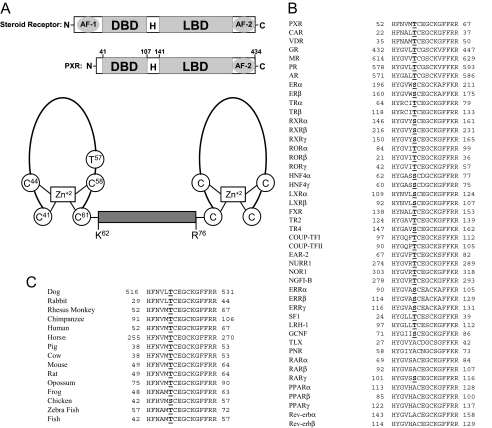
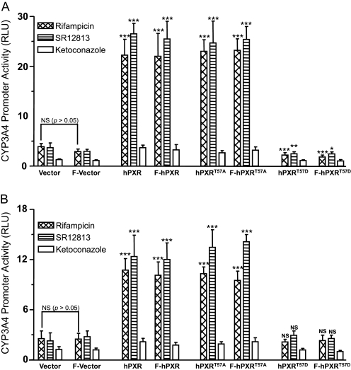
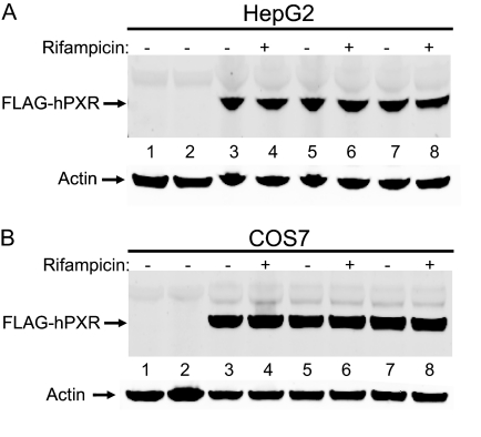
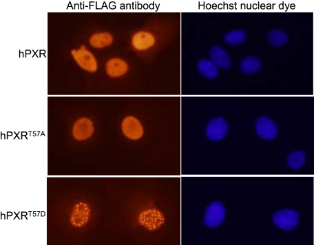
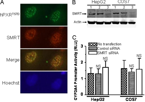
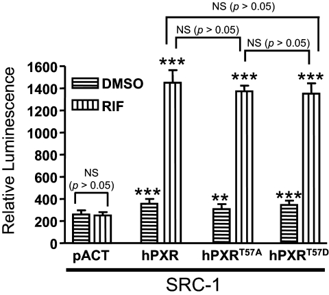
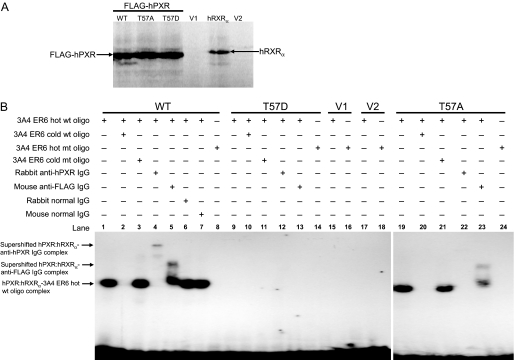
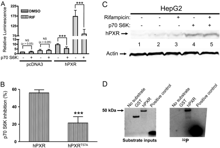
The pregnane X receptor (PXR) plays crucial roles in multiple physiological processes. However, the signaling mechanisms responsible are not well defined; it is most likely that multiple functions of PXR are modulated by its phosphorylation. Therefore, we sought to determine whether mutation at a highly conserved Thr(57) affects human PXR (hPXR) function. Site-directed mutagenesis was performed to generate phosphorylation-deficient (hPXR(T57A)) and phosphomimetic (hPXR(T57D)) mutants. Gene reporter, Western blotting, immunocytochemistry, mammalian two-hybrid, and electrophoretic mobility shift assays were used to study cytochrome P450 3A4 (CYP3A4) promoter activation, protein levels, localization, cofactor interaction, and CYP3A4 promoter binding of the hPXR mutants, respectively. hPXR(T57D), but not hPXR(T57A), lost its transcriptional activity. Neither mutation altered hPXR's protein levels and interaction with steroid receptor coactivator-1. hPXR and hPXR(T57A) exhibited a homogenous nuclear distribution, whereas hPXR(T57D) exhibited a distinctive punctate nuclear localization pattern similar to that of hPXR mutants with impaired function that colocalize with silencing mediator of retinoid and thyroid receptors (SMRT), although silencing of SMRT did not rescue the altered function of hPXR(T57D). However, hPXR(T57D), but not hPXR(T57A), impaired hPXR's ability to bind to the CYP3A4 promoter, consistent with the mutant's transactivation function. Furthermore, the 70-kDa form of ribosomal protein S6 kinase (p70 S6K) phosphorylated hPXR in vitro and inhibited its transcriptional activity, whereas hPXR(T57A) partially resisted the inhibitory effect of p70 S6K. Our studies identify a functionally significant phosphomimetic mutant (hPXR(T57D)) and show p70 S6K phosphorylation and regulation of hPXR transactivation to support the notion that phosphorylation plays important roles in regulating hPXR function.


Figures

F<sc>ig</sc>. 1.
Fig. 1.
Thr57 in hPXR is conserved in other NRs. A, top panel showing the schematic comparison of the domain structures of a steroid receptor and hPXR. H, hinge region. Bottom panel shows the schematic representation of the two zinc finger motifs in the DBD of hPXR. The two zinc finger motifs are separated by a linker (from Lys62 to Arg76). The diagram was not drawn as per the scale in terms of number of residues. Note that Thr57 is located within the first zinc finger motif. B, Thr57 in hPXR is highly conserved among other human NRs. Of the 46 human NRs with DBD, 37 receptors (including all the classic steroid receptors) have a conserved T/S in the DBD. Nine receptors are exceptions, and they have an A instead of Thr/Ser at this position. Expansion of the abbreviated nomenclature for the receptors is as follows: CAR, constitutive androstane receptor; VDR, vitamin D receptor; GR, glucocorticoid receptor; MR, mineralocorticoid receptor; PR, progesterone receptor; AR, androgen receptor; ER, estrogen receptor; TR, thyroid hormone receptor; ROR, retinoid-related orphan receptor; LXR, liver X receptor; FXR, farnesoid X receptor; TR2, testicular receptor 2; TR4, testicular receptor 4; COUP-TF, chicken ovalbumin upstream promoter transcription factor; EAR-2, v-erbA-related; NURR1, NR-related 1; NOR1, neuron-derived orphan receptor 1; NGFI-B, nerve growth factor-induced clone B; ERR, estrogen receptor-related; SF-1, steroidogenic factor 1; LRH-1, liver receptor homolog-1; GCNF, germ cell nuclear factor; TLX, human homolog of the Drosophila tailless gene; PNR, photoreceptor cell-specific NR. C, Thr57 in hPXR is conserved in all the vertebrate species with a fully known PXR sequence. Chicken PXR is the only exception and has an S instead of T at that position.
F<sc>ig</sc>. 2.
Fig. 2.
Phosphomimetic mutation at Thr57 abolishes hPXR transactivation activity. HepG2 (A) and COS7 (B) cells were cotransfected with either pcDNA3 or hPXR using CYP3A4-luc. F denotes for FLAG-tag. Twenty-four hours post-transfection, the cells were treated with vehicle DMSO (0.1%) or 10 μM compounds: rifampicin, SR12813, and ketoconazole. Luciferase activity was measured 24 h after compound treatments. CYP3A4 promoter activity induced by hPXR after treatment with the compounds was shown as RLUs. RLUs were determined by normalizing the luminescence observed in the presence of one of the compounds with the luminescence observed in the presence of DMSO. Data are shown as mean values from six independent experiments with bars indicating the S.D. The Student's t test was used to determine statistical significance of unpaired samples by comparing the RLU obtained from the samples transfected with wild-type or mutant hPXR with the samples transfected with the vector. Likewise, statistical significance was determined from the samples transfected with FLAG-tagged wild-type or mutant hPXR to the samples transfected with the FLAG vector. Furthermore, statistical significance was ascertained similarly by comparing the RLU obtained from the samples transfected with FLAG-tagged plasmids with the samples transfected with corresponding untagged plasmids, and statistics were shown only for one comparison, i.e., between the vector and FLAG vector after rifampicin treatment. No statistical significance was observed between the samples transfected with the FLAG-tagged plasmids and the untagged plasmids (statistics were not shown for all the sample comparisons). Differences were considered significant for p < 0.05 (*), 0.01 (**), or 0.001 (***), and nonsignificant (N.S.) for p > 0.05.
F<sc>ig</sc>. 3.
Fig. 3.
Mutation at Thr57 does not affect hPXR protein expression levels. HepG2 (A) and COS7 (B) cells were transfected with either wild-type or mutant FLAG-hPXR plasmids. Twenty-four hours post-transfection, the cells were treated with either 0.1% DMSO (indicated by a “–” symbol) or 10 μM rifampicin (indicated by a“+” symbol). Whole cell lysates were collected 24 h after treatment with DMSO or rifampicin and subjected to Western blotting analysis using anti-FLAG and anti-actin antibodies as described under Materials and Methods. pcDNA3-FLAG vector transfection (lane 1) and no transfection (lane 2) are “negative” controls. Wild-type FLAG-hPXR protein expression was shown in lanes 3 and 4. Protein expression for the alanine mutant (FLAG-hPXRT57A) was shown in lanes 5 and 6 and for the aspartate (FLAG-hPXRT57D) in lanes 7 and 8. Actin protein expression was analyzed as a mean of loading control. Data shown are from a representative experiment.
F<sc>ig</sc>. 4.
Fig. 4.
Phosphomimetic mutation at Thr57 alters hPXR nuclear localization pattern. COS7 cells were transfected with FLAG-hPXR, FLAG-hPXRT57A, or FLAG-hPXRT57D plasmids. The cells were treated with vehicle 0.1% DMSO or 10 μM rifampicin 24 h post-transfection. The cells were processed for immunocytochemistry 24 h after treatment with DMSO or rifampicin as described under Materials and Methods. Wild-type and mutant hPXR were probed with Cy3-conjugated anti-FLAG M2 mouse monoclonal antibody. Nuclear DNA was stained with Hoechst dye. Data shown are from a representative experiment.
F<sc>ig</sc>. 5.
Fig. 5.
Silencing of SMRT cannot rescue the impaired function of hPXRT57D. A, hPXRT57D colocalizes with SMRT to the nucleus at discrete nuclear foci. COS7 cells were transfected with FLAG-hPXRT57D. Twenty-four hours post-transfection, the cells were treated with vehicle 0.1% DMSO or 10 μM rifampicin. The cells were processed for immunocytochemistry 24 h after treatment with DMSO or rifampicin. FLAG-hPXRT57D was probed using rabbit anti-hPXR polyclonal antibody; SMRT was probed using anti-SMRT mouse monoclonal antibody; and nuclear DNA was stained with Hoechst dye. Data shown are from a representative experiment. B, Accell SMARTpool human SMRT siRNA knocks down the protein expression of SMRT. HepG2 and COS7 cells were transfected with 1 μM Accell nontargeting pool control siRNA (lane 2) or Accell SMARTpool human SMRT siRNA (lane 3) in Accell siRNA delivery media. Whole cell lysates were collected 72 h after transfection and subjected to Western blotting analysis using anti-SMRT and anti-actin antibodies. Protein expression for SMRT without transfection was shown in lane 1. Actin protein expression was shown as a loading control. Data shown are from a representative experiment. C, silencing of SMRT cannot rescue the impaired transactivation function of hPXRT57D. HepG2 and COS7 cells were transfected with pcDNA3-FLAG-PXRT57D, pGL4-hRluc, and pGL3-CYP3A4-luc using Lipofectamine. Five hours after transfection with the plasmids, the cells were transfected with 1 μM Accell nontargeting SMARTpool control siRNA or Accell SMARTpool SMRT siRNA in Accell siRNA delivery media for 72 h. Forty-eight hours after transfection with the siRNAs, the cells were treated with vehicle DMSO (0.1%) or 10 μM rifampicin. Firefly and Renilla luciferase activities were measured 24 h after rifampicin treatment. Firefly luciferase activity was first normalized with Renilla luciferase activity before normalizing the data for rifampicin with DMSO. Data are shown as mean values from four independent experiments with bars indicating the S.D. The Student's t test was used to determine statistical significance of unpaired samples by comparing the RLU obtained from the samples transfected with control siRNA or SMRT siRNA with the samples that were not transfected with siRNA. Differences were considered nonsignificant (N.S.) for p > 0.05.
F<sc>ig</sc>. 6.
Fig. 6.
Phosphomimetic mutation at Thr57 does not affect hPXR interaction with SRC-1. Mammalian two-hybrid assays were performed in HepG2 cells transiently cotransfected with plasmids encoding Gal4-SRC-1 and the reporter gene pG5-luc, along with empty vector pACT, VP16-hPXR, VP16-hPXRT57A, or VP16-hPXRT57D, as indicated. The cells were treated with DMSO or 5 μM rifampicin (RIF) 24 h after transfection. Luciferase assays were performed 24 h after the compound treatment. The relative luminescence for pG5-luc was determined by normalizing firefly luciferase activity with Renilla luciferase activity. The values represent the means of eight independent experiments, and the bars denote the S.D. The Student's t test was used to determine statistical significance of unpaired samples by comparing the relative luminescence obtained between the samples cotransfected with the vector and hPXR plasmids for corresponding DMSO or rifampicin treatments. Comparisons between other samples were noted in brackets. Differences were considered significant for p < 0.05 (*), 0.01 (**), or 0.001 (***), and nonsignificant (N.S.) for p > 0.05. SRC-1, Gal4-SRC-1; hPXR, VP16-hPXR; hPXRT57A, VP16-hPXRT57A; hPXRT57D, VP16-hPXRT57D.
F<sc>ig</sc>. 7.
Fig. 7.
Phosphomimetic mutation at Thr57 impairs DNA binding of hPXR. A, in vitro synthesis of proteins for wild-type and mutant FLAG-hPXR, as well as for hRXRα, as described under Materials and Methods. Equal amounts of protein were observed from 2-μl in vitro transcription and translation reactions for wild-type (WT), alanine mutant (T57A), and aspartate mutant (T57D) hPXRs. No specific proteins were observed from the negative control reactions using the vectors, pcDNA3-FLAG (V1) and pSG5 (V2), for hPXR and hRXRα, respectively. B, electrophoretic mobility shift assay was performed with in vitro-translated FLAG-hPXR, FLAG-hPXRT57D, or FLAG-hPXRT57A and hRXRα proteins, as well as 32P-labeled CYP3A4 PXR DNA binding sequence (indicated as 3A4ER6) as described under Materials and Methods. Equal amount of hRXRα was added to all the reactions and was not shown in the figure. FLAG-hPXR and FLAG-hPXRT57A bound to and formed a complex with hot wild-type (wt) oligo, and this complex (lanes 1 and 19) was efficiently competed with by cold wt oligo (lanes 2 and 20) but not by cold mutant (mt) oligo (lanes 3 and 21) at 500 M excess. Addition of 2 μg of rabbit anti-hPXR IgG supershifted the FLAG-hPXR-hot wt oligo complex (lane 4) and vanished the FLAG-hPXRT57A-hot wt oligo complex (lane 22) (Saradhi et al., 2005). Addition of 2 μg of mouse anti-FLAG IgG supershifted both the FLAG-hPXR-hot wt oligo complex (lane 5) and the FLAG-hPXRT57A-hot wt oligo complex (lane 23). Addition of 2 μg of rabbit (lane 6) and mouse (lane 7) normal IgG failed to supershift or vanish the FLAG-hPXR-hot wt oligo complex. No complex was detected for FLAG-hPXR (lane 8) and FLAG-hPXRT57A (lane 24) with hot mt oligo. No complex was detected with phosphomimetic mutant FLAG-hPXRT57D (lanes 9–14) and vectors, pcDNA3-FLAG (V1) (lanes 15 and 16), and pSG5 (V2) (lanes 17 and 18). Data shown are from a representative experiment.
F<sc>ig</sc>. 8.
Fig. 8.
p70 S6K regulates hPXR function. A, p70 S6K attenuates the transactivating activity of hPXR. HepG2 cells were cotransfected with pGL4-hRluc and pGL3-CYP3A4-luc, along with either FLAG-pcDNA3, pcDNA3-FLAG-hPXR, or pcDNA3-FLAG-hPXRT57A (for B), with or without the constitutively active p70 S6K, as indicated. Twenty-four hours after transfection, the cells were treated with vehicle DMSO (0.01%) or 1 μM rifampicin (RIF). Firefly and Renilla luciferase activities were measured 24 h after rifampicin treatment. CYP3A4 promoter activity was shown as relative luminescence determined by normalizing the firefly luciferase activity with Renilla luciferase activity. Data are shown as mean values from four to six independent experiments, with bars indicating the S.D. The Student's t test was used to determine statistical significance of unpaired samples by comparing the relative luminescence between samples as noted in brackets. Differences were considered significant for p < 0.05 (*), 0.01 (**), or 0.001 (***), and nonsignificant (N.S.) for p > 0.05. pcDNA3, FLAG-pcDNA3; hPXR, pcDNA3-FLAG-hPXR. B, hPXRT57A partially resists the regulation of p70 S6K. Percentage inhibition of rifampicin-inducible hPXR-transactivating activity by p70 S6K was shown. The change (inhibition) in rifampicin-inducible hPXR activity after p70 S6K cotransfection, for the corresponding wild-type or mutant hPXR, was calculated and expressed as percentage inhibition. Data are shown as mean values from four to six independent experiments, with bars indicating the S.D. The Student's t test was used to determine statistical significance of unpaired samples. Differences were considered significant for p < 0.05 (*), 0.01 (**), or 0.001 (***). hPXR, pcDNA3-FLAG-hPXR; hPXRT57A, pcDNA3-FLAG-hPXRT57A. C, p70 S6K cotransfection does not reduce hPXR protein levels. Transient transfections were performed as described in A. Whole cell lysates were collected 24 h after treatment with DMSO (0.01%) or 1 μM rifampicin and subjected to Western blotting analysis using rabbit polyclonal anti-hPXR serum (Saradhi et al., 2005). FLAG-pcDNA3 vector transfection (lane 1) is a negative control. pcDNA3-FLAG-hPXR protein expression without p70 S6K cotransfection was shown in lanes 2 and 3, and with p70 S6K cotransfection was shown in lanes 4 and 5. Actin protein expression was analyzed as a mean of loading control. D, p70 S6K phosphorylates hPXR in vitro. Right, recombinant active p70 S6K (20 ng) was used with 1 μg of substrate as indicated. The kinase assays were performed as described under Materials and Methods. Left, SimplyBlue staining of the proteins resolved on SDS-polyacrylamide gel electrophoresis indicating the amounts of input substrate proteins used in the kinase assay reactions.

Similar articles

Cited by

References

    1. Bauer B, Yang X, Hartz AM, Olson ER, Zhao R, Kalvass JC, Pollack GM, and Miller DS (2006) In vivo activation of human pregnane X receptor tightens the blood-brain barrier to methadone through P-glycoprotein up-regulation. Mol Pharmacol 70 1212–1219. - PubMed
    1. Bhalla S, Ozalp C, Fang S, Xiang L, and Kemper JK (2004) Ligand-activated pregnane X receptor interferes with HNF-4 signaling by targeting a common coactivator PGC-1alpha. Functional implications in hepatic cholesterol and glucose metabolism. J Biol Chem 279 45139–45147. - PubMed
    1. Chen D, Pace PE, Coombes RC, and Ali S (1999) Phosphorylation of human estrogen receptor alpha by protein kinase A regulates dimerization. Mol Cell Biol 19 1002–1015. - PMC - PubMed
    1. Chen Y, Tang Y, Wang MT, Zeng S, and Nie D (2007) Human pregnane X receptor and resistance to chemotherapy in prostate cancer. Cancer Res 67 10361–10367. - PubMed
    1. Cohen P (2000) The regulation of protein function by multisite phosphorylation—a 25 year update. Trends Biochem Sci 25 596–601. - PubMed

LinkOut - more resources