. 2009 Jan 26;184(2):215-23.
doi: 10.1083/jcb.200808080.

TREM-2 (triggering receptor expressed on myeloid cells 2) is a phagocytic receptor for bacteria

Affiliations

TREM-2 (triggering receptor expressed on myeloid cells 2) is a phagocytic receptor for bacteria

Elsa-Noah N'Diaye et al. J Cell Biol. .

Abstract

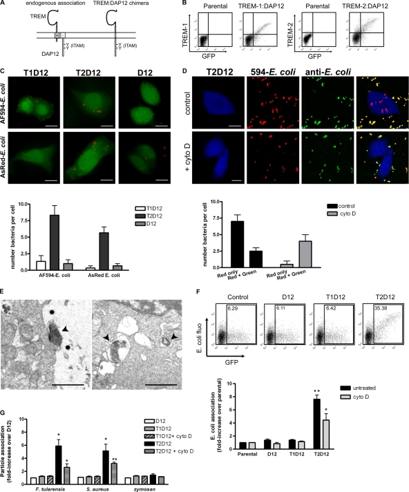
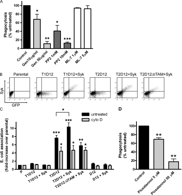
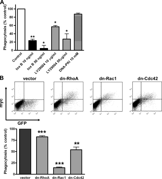
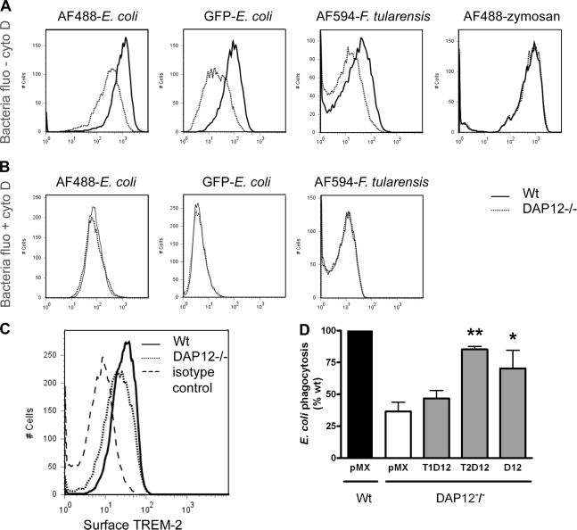
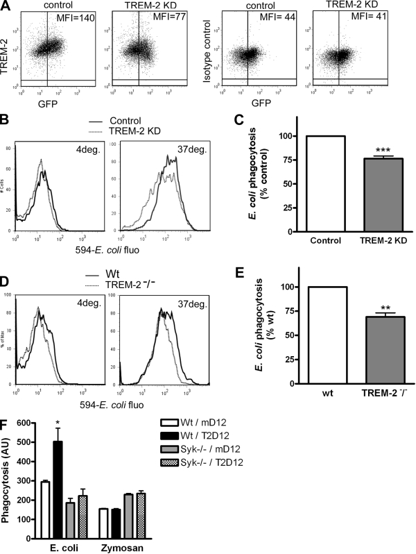
Phagocytosis, which is essential for the immune response to pathogens, is initiated by specific interactions between pathogens and cell surface receptors expressed by phagocytes. This study identifies triggering receptor expressed on myeloid cells 2 (TREM-2) and its signaling counterpart DAP12 as a molecular complex that promotes phagocytosis of bacteria. Expression of TREM-2-DAP12 enables nonphagocytic Chinese hamster ovary cells to internalize bacteria. This function depends on actin cytoskeleton dynamics and the activity of the small guanosine triphosphatases Rac and Cdc42. Internalization also requires src kinase activity and tyrosine phosphorylation. In bone marrow-derived macrophages, phagocytosis is decreased in the absence of DAP12 and can be restored by expression of TREM-2-DAP12. Depletion of TREM-2 inhibits both binding and uptake of bacteria. Finally, TREM-2-dependent phagocytosis is impaired in Syk-deficient macrophages. This study highlights a novel role for TREM-2-DAP12 in the immune response to bacterial pathogens.


Figures

Figure 1.
Figure 1.
Expression of TREM-2–DAP12 promotes binding and internalization of bacteria by nonphagocytic cells. (A) Schematic of TREM association with DAP12 in trans (endogenous conformation, left) or through covalent linkage (chimeric construct, right). (B) CHO cells were transfected with bicistronic constructs containing GFP together with TREM-1–DAP12 (T1D12) or TREM-2–DAP12 (T2D12) or DAP12 only (D12). Surface expression of TREM–DAP12 chimeras in GFP-positive cells was verified by flow cytometry using antibodies to TREM-1 or TREM-2. (C) T1D12, T2D12, or D12 cells were challenged with 594–E. coli (top) or AsRed–E. coli (bottom). Association of transfected cells with bacteria was analyzed by fluorescence microscopy and quantified (graph). (D) T2D12 cells challenged with 594–E. coli were stained with E. coli antibodies and analyzed by microscopy. Pseudocolors: blue, T2D12 cells; red, 594–E. coli; green, anti–E. coli staining. Internal bacteria are red, whereas extracellular bacteria are both red and green (yellow on merge). Both internalized and extracellular cell-bound bacteria were scored (graph). Arrowheads represent cell-bound bacteria. (E) T2D12 cells challenged with live, unlabeled E. coli were analyzed by electron microscopy. Both binding (left) and internalization of bacteria within phagosomes (right) were observed. Arrowheads, bacteria; asterisks, pseudopods. (F) Association of T2D12 cells with 594–E. coli was analyzed by flow cytometry and quantified by measuring the fluorescence intensity of bacteria associated with GFP-positive cells. Representative plots are shown. Internalization of bacteria (but not binding) was blocked with cyto D. Fluorescence intensities were normalized to values of parental cells. The graph represents the mean of four experiments ± SEM. *, P < 0.05; **, P < 0.01 relative to parental values. (G) T1D12, T2D12, or D12 cells were challenged with F. tularensis, S. aureus, or zymosan, and their association was quantified as in F. Fluorescence intensities were normalized to D12 values in each set. The graph represents the mean of three experiments ± SEM. *, P < 0.05; **, P < 0.01 relative to D12 values. Bars: (C and D) 10 µm; (E) 1 µm.
Figure 2.
Figure 2.
TREM-2–DAP12-mediated phagocytosis requires tyrosine phosphorylation and src kinase activity, and it is enhanced by Syk. (A) T2D12 cells were treated with genistein (tyrosine kinase inhibitor), PP2 (src kinase inhibitor), or ML-7 (myosin light chain kinase inhibitor) for 2 h at the indicated concentrations before addition of 594–E. coli. Controls for binding were treated with cyto D. Bacteria association was assessed by flow cytometry as in Fig 1. To quantify uptake, values obtained with cyto D were subtracted from that of untreated samples. The graph shows values normalized to controls (no inhibitor). *, P ≤ 0.05; **, P < 0.01; ***, P < 0.001 relative to controls. (B) CHO cells were transfected with T1D12, T2D12, D12, or a variant of T2D12 with a mutated ITAM (T2D12ΔITAM). Where indicated, cells were cotransfected with Syk. (C) 594–E. coli association with transfected cells was quantified as in Fig. 1 F. *, P ≤ 0.05; **, P < 0.01; ***, P < 0.001 relative to parental values. (D) T2D12 cells treated with the indicated concentrations of piceatannol were challenged with E. coli, and uptake was analyzed as in Fig. 2 A. **, P < 0.01. The graphs represent the mean of three experiments ± SEM.
Figure 3.
Figure 3.
TREM-2–DAP12-mediated phagocytosis requires Rac1 and Cdc42 activity. (A) T2D12 cells were treated with C. difficile toxin B (Rho GTPase family inhibitor), LY29004 (PI-3 kinase inhibitor), or DNA–protein kinase inhibitor before phagocytosis of 594–E. coli. *, P < 0.05; **, P < 0.01 relative to untreated conditions. (B) Expression of dn Rho, Rac, or Cdc42 into T2D12 cells was verified by flow cytometry. Phagocytosis was analyzed as in A. **, P < 0.01; ***, P < 0.001 relative to vector-transfected cells. The graphs represent the mean of three experiments ± SEM.
Figure 4.
Figure 4.
Expression of TREM-2–DAP12 restores phagocytosis in DAP12−/− macrophages. (A and B) wt and DAP12−/− BMDMs were challenged with E. coli (surface or internally labeled), F. tularensis, and zymosan. Particle association was assessed by flow cytometry in the absence (A) or in the presence of cyto D or at 4°C (B). Representative histograms are shown. (C) Surface expression of TREM-2 was analyzed on wt and DAP12−/− BMDMs. (D) DAP12−/− BMDMs were transduced with DAP12, TREM-1–DAP12, TREM-2–DAP12, or empty vector (pMX). Phagocytosis of 594–E. coli by transduced cells was compared with that of wt BMDMs transduced with empty vector (100%). The graph represents the mean of four experiments ± SEM. *, P ≤ 0.05; **, P < 0.01 relative to DAP12−/−/pMX values.
Figure 5.
Figure 5.
TREM-2 promotes phagocytosis of bacteria by macrophages in a Syk-dependent manner. (A) Cellular levels of TREM-2 on BMDMs were decreased by the expression of a short hairpin RNA (TREM-2 KD) and measured by flow cytometry. An isotype antibody was used as a negative control. MFI, mean fluorescence intensity. (B) E. coli association with control or TREM-2 KD BMDMs at 4 or 37°C was assessed by flow cytometry. (C) Bacteria uptake was quantified by subtracting 4°C from 37°C fluorescence values. The graph represents the mean of five experiments ± SEM. ***, P ≤ 0.001. (D and E) E. coli association with wt or TREM-2−/− BMDMs was assessed as in B, and bacteria uptake was quantified in E as described in C. The graph represents the mean of three experiments + SEM. **, P ≤ 0.01. (F) TREM-2–D12 or DAP12 alone (mutated in its transmembrane domain) was expressed in wt or Syk−/− BMDMs, and phagocytosis of E. coli or zymosan was quantified by subtracting 4°C from 37°C fluorescence values. The graph represents the mean of three experiments ± SEM. *, P ≤ 0.05 between wt/mutated DAP12 and wt/TREM-2–DAP12. AU, arbitrary unit.

References

    1. Aderem A., Underhill D.M. 1999. Mechanisms of phagocytosis in macrophages.Annu. Rev. Immunol. 17:593–623 - PubMed
    1. Andrews S., Stephens L.R., Hawkins P.T. 2007. PI3K class IB pathway in neutrophils.Sci. STKE. doi:10.1126/stke.4072007cm3 - DOI - PubMed
    1. Bakker A.B., Hoek R.M., Cerwenka A., Blom B., Lucian L., McNeil T., Murray R., Phillips L.H., Sedgwick J.D., Lanier L.L. 2000. DAP12-deficient mice fail to develop autoimmunity due to impaired antigen priming.Immunity. 13:345–353 - PubMed
    1. Berton G., Mocsai A., Lowell C.A. 2005. Src and Syk kinases: key regulators of phagocytic cell activation.Trends Immunol. 26:208–214 - PubMed
    1. Brown E.J. 1995. Phagocytosis.Bioessays. 17:109–117 - PubMed

Publication types