. 2009 Jan 26;184(2):225-39.
doi: 10.1083/jcb.200808049.

The SCF Slimb ubiquitin ligase regulates Plk4/Sak levels to block centriole reduplication

Affiliations

The SCF Slimb ubiquitin ligase regulates Plk4/Sak levels to block centriole reduplication

Gregory C Rogers et al. J Cell Biol. .

Abstract

Restricting centriole duplication to once per cell cycle is critical for chromosome segregation and genomic stability, but the mechanisms underlying this block to reduplication are unclear. Genetic analyses have suggested an involvement for Skp/Cullin/F box (SCF)-class ubiquitin ligases in this process. In this study, we describe a mechanism to prevent centriole reduplication in Drosophila melanogaster whereby the SCF E3 ubiquitin ligase in complex with the F-box protein Slimb mediates proteolytic degradation of the centrosomal regulatory kinase Plk4. We identified SCF(Slimb) as a regulator of centriole duplication via an RNA interference (RNAi) screen of Cullin-based ubiquitin ligases. We found that Plk4 binds to Slimb and is an SCF(Slimb) target. Both Slimb and Plk4 localize to centrioles, with Plk4 levels highest at mitosis and absent during S phase. Using a Plk4 Slimb-binding mutant and Slimb RNAi, we show that Slimb regulates Plk4 localization to centrioles during interphase, thus regulating centriole number and ensuring the block to centriole reduplication.


Figures
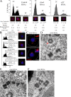
Figure 1.
Figure 1.
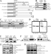
slimb RNAi elevates centriole number. (A) Effects of slimb RNAi are not solely via effects on cell cycle. Cells stained for D-PLP (red) and DNA (blue) were quantitated for DNA content by HTM (5,000 cells/histogram). Centrioles were manually counted in ∼150 randomly selected cells with 2C, 4C, or 8C DNA. Example cells and mean, median, and range of centriole numbers are shown below sampled histogram regions. Histograms are not shown to scale. Asterisks denote a significant difference; *, P < 0.007 (unpaired t test). Slimb or E2F1 depletion eliminates the 8C population. (B) Cell cycle progression (assessed via HTM; 5,000 cells/histogram) and centriole number were scored in day 7 RNAi-treated cells. D-PLP–labeled centrioles (red) were manually counted. Histograms are shown to scale. (C) Unique centriole configurations in Slimb-depleted cells that contained more than the mean number of D-PLP–labeled centrioles (red). White tracing marks cell borders. Insets show centrioles at a higher magnification. (A–C) Bars, 5 µm. (D–F) Transmission electron micrographs of interphase Slimb-depleted cells. (D) Typical centriolar microtubule arrangement of the centriole in cross section (red arrow). Other centrioles are sagittal sections oriented in different manners. Excess centrioles in rows (E) and clusters (F).
Figure 2.
Figure 2.
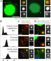
Slimb localizes to centrioles. (A) Immunoblot of affinity-purified anti-Slimb antibody against an S2 cell lysate. (B) slimb RNAi depletes protein by 98%. (C and C′) Immunostaining of Slimb (green, monochrome) and D-PLP centrioles (red) in interphase S2 cells. (D and D′) Stable S2 line expressing Slimb-EGFP (green, monochrome) and mCherry–SAS-6 centrioles in live interphase cells. (C–D′) Centrioles (arrowheads) are shown at a higher magnification (insets). (E) Endogenous Slimb and Slimb-EGFP cosediment with centrioles purified from S2 cells on a 20–70% sucrose gradient. Fractions were immunoblotted for D-PLP, GFP, and Slimb. Asterisks denote peak centriole-containing fractions. (F) Slimb (green, arrowheads) immunolocalizes to D-PLP centrioles (red) after 24-h drug-induced S-, G2-, or M-phase arrest. Histograms (to scale) of DNA content assessed by HTM (5,000 cells/histogram). Condensed DNA (blue) reveals a mitotic cell. Insets show centrioles at a higher magnification. (G) Graph of endogenous Slimb levels after 24-h drug-induced cell cycle arrest as determined from quantitative anti-Slimb immunoblots (below graph). (B and G) α-Tubulin was used as a loading control. Bars, 5 µm.
Figure 3.
Figure 3.
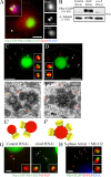
Plk4 is degraded in a Slimb-dependent manner and is stabilized by perturbing its interaction with Slimb. (A) Plk4 family showing the conserved kinase domain (gray), Polo box motif (striped boxes), and Slimb-binding consensus (black bars). The S293A/T297A SBM should be nondegradable. (B) Preimmune control and anti-Slimb immunoprecipitates from stable S2 cell lysates expressing Plk4-myc were probed for anti-Slimb, SkpA, and myc. (C) Control and anti-GFP immunoprecipitates from S2 cell lysates transiently expressing either inducible wild-type Plk4-EGFP or Plk4-SBM–EGFP were probed for endogenous Slimb. IP, immunoprecipitation. (D) Anti-GFP immunoprecipitates from S2 cell lysates transiently expressing triple Flag-ubiquitin and either inducible wild-type Plk4-EGFP or Plk4-SBM–EGFP were probed with anti-GFP (left) and anti-Flag (right) antibody. IB, immunoblot. (E) Anti-GFP immunoblots of lysates prepared from stable SAS-6p–driven Plk4-EGFP that were RNAi treated for the indicated protein for 7 d. (F) Plk4 is phosphorylated. (left) Anti-GFP immunoblots of lysates from control or Slimb-depleted cells transiently expressing inducible Plk4-EGFP. Plk4 accumulates as a doublet (arrowheads) after slimb RNAi. (right) Anti-GFP immunoprecipitates from day 4 Slimb-depleted cells transiently expressing inducible Plk4-EGFP were mock or alkaline phosphatase treated. Plk4 shifts from a broad band (bracket) to a faster migrating single polypeptide (arrowhead). (D–F) Molecular mass is indicated in kilodaltons. (G) Anti-GFP immunoblots showing the levels of transiently expressed SAS-6p–driven Plk4-EGFP and Plk4-SBM–EGFP in 24-h drug-induced cell cycle–arrested cells. Cotransfected Nlp-EGFP was used as a loading control. (H) Anti-GFP immunoblots showing the levels of 4 h–induced Plk4-EGFP and Plk4-SBM–EGFP expression in drug-induced cell cycle–arrested cells. (E and H) α-Tubulin was used as a loading control.
Figure 4.
Figure 4.
Slimb regulates Plk4 levels on centrioles to control centriole number. (A) Asymmetrical Plk4 localization. Transient coexpression of Nlp-EGFP (Ito et al., 1996) as a cotransfection marker (green) labeling nuclei (arrow) and Plk4-EGFP (green). D-PLP (red) marks centrioles (arrowheads). Insets show centrioles at a higher magnification. (B–D) Mutating the Slimb-binding site stabilizes Plk4 on centrioles. (B) Cell cycle distributions after 24-h drug-induced S, G2, or mitotic arrest. Histograms are shown to scale and were assessed by HTM (5,000 cells/histogram). (C) Plk4-EGFP (green) only localizes to M-phase centrioles (arrowheads and insets) marked with mCherry–SAS-6 in live cells (red). Condensed DNA (blue) reveals mitotic cells. (D) Plk4-SBM–EGFP (green) localizes to centrioles during all cell cycle phases that were examined. Centrioles (arrowheads and insets) are marked with D-PLP in fixed cells (red). Nlp-EGFP (green nuclei, cytoplasmic during mitosis) is the cotransfection control. Bars: (A) 5 µm; (C and D) 2.5 µm.
Figure 5.
Figure 5.
Stable Plk4 promotes excess daughter centriole formation, and slimb RNAi eliminates the S-phase centriole reduplication block by accumulating Plk4 on centrioles. (A) Slimb overlaps Plk4-SBM–EGFP localization on centrioles. Immunostaining of Slimb (red) and D-PLP centrioles (blue) in a transiently expressing coexpressing Nlp-EGFP (green nuclei) and SAS-6p Plk4-SBM–EGFP (green) interphase S2 cell. A representative centriole (arrowhead) is shown at a higher magnification (inset). (B) Anti-GFP immunoblots of lysates prepared from transiently expressing inducible Plk4-EGFP that were RNAi treated for the indicated proteins for 7 d. α-Tubulin was used as a loading control. Molecular mass is indicated in kilodaltons. (C and D) Transient coexpression of Nlp-EGFP (green nuclei) and Plk4-SBM–EGFP in day 5 cycling S2 cells. Plk4-SBM–EGFP labels one or more spots (green) on D-PLP–stained centrioles (red). Insets show select centrioles at a higher magnification. The cell in D shows an extreme example of centriole overduplication. White tracing marks cell borders. (E and F) Transmission electron micrographs of interphase cells expressing Plk4-SBM–EGFP for 5 d. Red arrows denote excess daughter centrioles emanating from mother centrioles shown in the cross section. The cell in E shows a normal mother–daughter centriole pair (orange arrow) adjacent to a mother with two daughters. Illustrations of these centrioles are shown in E′ and F′. (G) Transient expression of Plk4-EGFP/Nlp-EGFP (green) in day 3 RNAi-treated cells arrested in S phase for 2 d. D-PLP–labeled centrioles (red, arrowheads). Insets show centrioles at a higher magnification. (H) Stable expression of Plk4-EGFP (green) in a 24-h S phase–arrested cell treated with MG132 proteasome inhibitor. Insets show select D-PLP–labeled centrioles (red) at a higher magnification. Bars: (A–D) 5 µm; (E and F) 0.2 µm; (G and H) 2.5 µm.
Figure 6.
Figure 6.
Speculative model for a mechanism to limit centriole duplication to once per cell cycle by modulating the levels of Plk4 on centrioles through the activity of the SCFSlimb ubiquitin ligase. (1) Plk4 levels on centrioles peak during mitosis but also appear asymmetrically positioned on centrioles in a subpopulation of interphase cells. At this time, Plk4 activity initiates the duplication process by ”priming” centrioles for the duplication event that occurs later in the cell cycle. This could be achieved by targeting or stabilizing a key centriolar subunit to the parent centriole that then lays the foundation to assemble a procentriole. During mitotic exit, centriole pairs separate (disengage), thereby releasing centriole singlets into the interphase cytoplasm (Callaini and Riparbelli, 1990). Although Slimb localizes to centrioles during all of the cell cycle phases that we examined, Plk4 is not phosphorylated on residues required for Slimb binding during mitosis and is thus stable. (2) As cells complete cytokinesis, centriole singlets shed their PCM and lack microtubule nucleating activity (Rogers et al., 2008). During interphase, Plk4 is phosphorylated and now recognized by SCFSlimb, leading to its ubiquitination and degradation. Levels of centriole-associated Plk4 are low at this time. However, centrioles retain a critical modification (shown in purple) endowed upon them by Plk4 and are competent to duplicate. We note that Slimb and Plk4 levels on centrioles have not been determined during G1 phase. (3) Centrioles duplicate just before or during S phase with the appearance of a procentriole. Slimb on centrioles ensures that Plk4 levels remain low at this time and thus block centriole reduplication. (4) During G2, daughter centrioles elongate. Slimb at centrioles continues to prevent Plk4 accumulation.

References

    1. Balczon R., Bao L., Zimmer W.E., Brown K., Zinkowski R.P., Brinkley B.R. 1995. Dissociation of centrosome replication events from cycles of DNA synthesis and mitotic division in hydroxyurea-arrested Chinese hamster ovary cells.J. Cell Biol. 130:105–115 - PMC - PubMed
    1. Bettencourt-Dias M., Rodrigues-Martins A., Carpenter L., Riparbelli M., Lehmann L., Gatt M.K., Carmo N., Balloux F., Callaini G., Glover D.M. 2005. SAK/PLK4 is required for centriole duplication and flagella development.Curr. Biol. 15:2199–2207 - PubMed
    1. Blow J.J., Dutta A. 2005. Preventing re-replication of chromosomal DNA.Nat. Rev. Mol. Cell Biol. 6:476–486 - PMC - PubMed
    1. Boveri T. 1929. The Origin of Malignant Tumors.Williams and Wilkins Co., Baltimore, MD: 119 pp
    1. Brinkley B.R. 2001. Managing the centrosome numbers game: from chaos to stability in cancer cell division.Trends Cell Biol. 11:18–21 - PubMed

Publication types

MeSH terms