. 2009 Feb 16;206(2):387-98.
doi: 10.1084/jem.20070723. Epub 2009 Jan 26.

Antibody to the dendritic cell surface activation antigen CD83 prevents acute graft-versus-host disease

Affiliations

Antibody to the dendritic cell surface activation antigen CD83 prevents acute graft-versus-host disease

John Wilson et al. J Exp Med. .

Erratum in

  • J Exp Med. 2009 May 11;206(5):1203

Abstract

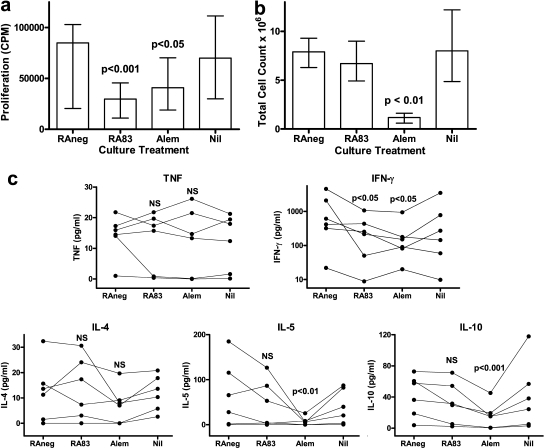
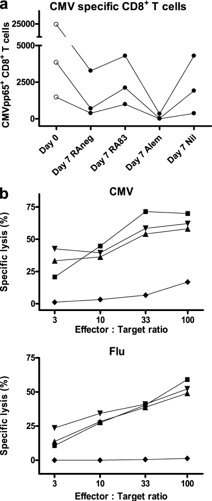
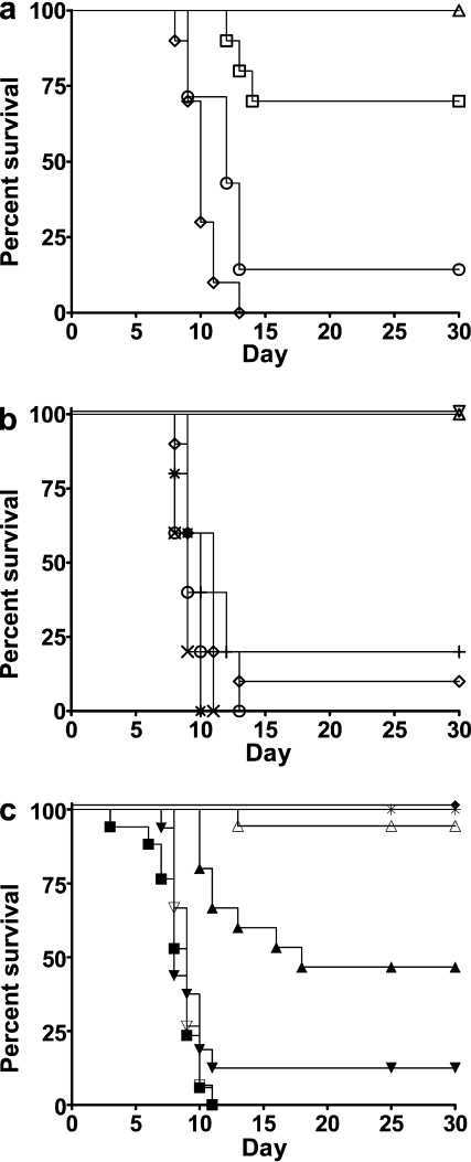
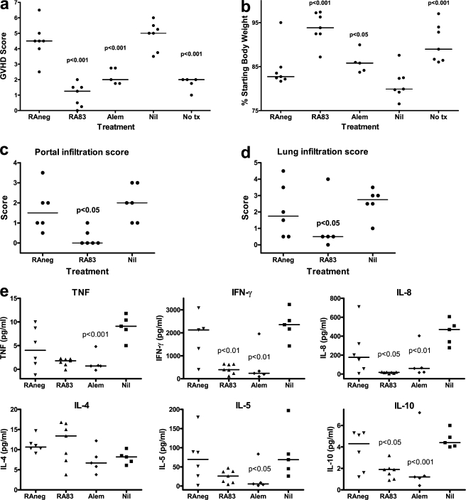
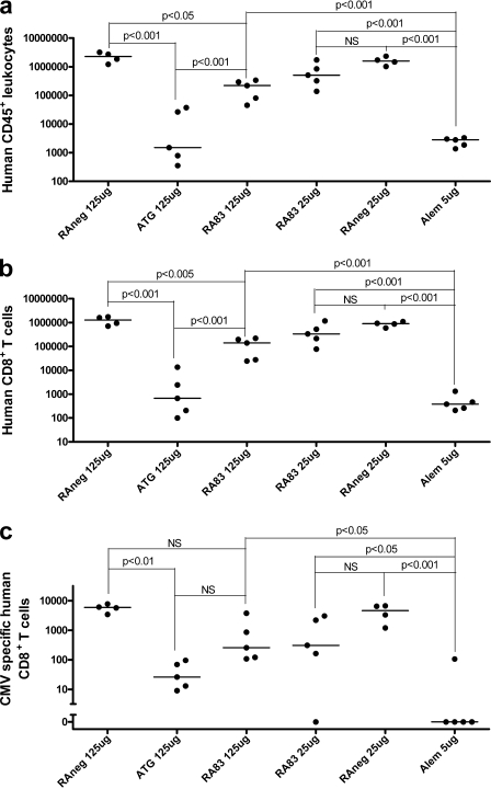
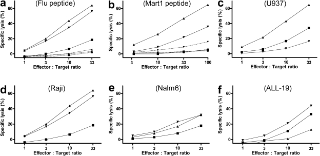
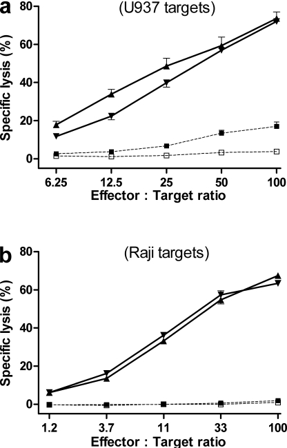
Allogeneic (allo) hematopoietic stem cell transplantation is an effective therapy for hematological malignancies but it is limited by acute graft-versus-host disease (GVHD). Dendritic cells (DC) play a major role in the allo T cell stimulation causing GVHD. Current immunosuppressive measures to control GVHD target T cells but compromise posttransplant immunity in the patient, particularly to cytomegalovirus (CMV) and residual malignant cells. We showed that treatment of allo mixed lymphocyte cultures with activated human DC-depleting CD83 antibody suppressed alloproliferation but preserved T cell numbers, including those specific for CMV. We also tested CD83 antibody in the human T cell-dependent peripheral blood mononuclear cell transplanted SCID (hu-SCID) mouse model of GVHD. We showed that this model requires human DC and that CD83 antibody treatment prevented GVHD but, unlike conventional immunosuppressants, did not prevent engraftment of human T cells, including cytotoxic T lymphocytes (CTL) responsive to viruses and malignant cells. Immunization of CD83 antibody-treated hu-SCID mice with irradiated human leukemic cell lines induced allo antileukemic CTL effectors in vivo that lysed (51)Cr-labeled leukemic target cells in vitro without further stimulation. Antibodies that target activated DC are a promising new therapeutic approach to the control of GVHD.


Figures

Figure 1.
Figure 1.
RA83 reduces T cell proliferation and expression of IFN-γ in allo MLC without nonspecific ablation of leukocytes. (a) Cell proliferation ([3H]thymidine incorporation; CPM) was significantly reduced in MLCs treated with 5 µg/ml of RA83 or with 5 µg/ml of alemtuzumab (Alem), compared with 5 µg/ml of RAneg (nonimmune rabbit IgG-negative control antibody). Median and interquartile range (error bars) are shown for n = 9 stimulator/responder combinations. (b) The number of viable leukocytes recovered from 7-d MLCs was not affected by RA83 but was substantially reduced by alemtuzumab. Median viable cell count and interquartile range (error bars) are shown for n = 11 stimulator/responder combinations. (c) RA83 reduced 7-d MLC concentrations of IFN-γ (note log scale) by a median of 64%, but TNF, IL-4, IL-5, and IL-10 were not significantly affected. Alemtuzumab similarly reduced 7-d MLC concentrations of IFN-γ (median 75% reduction), IL-5, and IL-10. Graphs show, for each antibody treatment, individual cytokine concentrations for n = 6 stimulator/responder combinations, each linked by lines. Raw cytokine data contained zero values; therefore, 1.0 pg/ml was added to all cytokine data to enable log transformation for statistical analysis (P > 0.05). NS and p-values are for repeated measures ANOVA followed by Bonferroni-corrected multiple comparisons posttests for RA83 and alemtuzumab each compared with RAneg treatment. Data are also shown for untreated (Nil) MLCs, which were not statistically significantly different from RAneg-treated cultures.
Figure 2.
Figure 2.
RA83 treatment preserves virus-specific T cell immunity in allo MLC. (a) Number of CMVpp65 pentamer-positive CD8+ T cells surviving after 7 d in antibody-treated 10-ml MLCs. The day 0 column shows the starting number of cells (data shown is for three different HLA-A*0201+ CMV+ donors; lines link data from the same donor). (b) A substantial viral antigen-specific functional CTL response was generated from 7-d RA83- (▴), RAneg- (▾), and Nil (▪)-treated MLCs but not from alemtuzumab (♦)-treated MLCs. Graphs show the mean percentage of lysis of CMV and FMP peptide-loaded 51Cr-labeled target cells by CTL effectors generated from the treated MLCs. P < 0.0001 for CMV (n = 3 donors) and P < 0.02 for FMP (n = 4 donors) for repeated measures ANOVA. Subsequent Bonferroni-corrected multiple comparisons testing showed that alemtuzumab treatment was significantly different from each of the other treatments (P < 0.001 for CMV and P < 0.01 for FMP).
Figure 3.
Figure 3.
Hu-SCID model of GVHD. (a) Human DC enable full GVHD induction. Administration of purified T cells (97% CD3+) alone induced severe GVHD in only 3 out of 10 mice (□; not significantly different from untransplanted controls [▵]), but coadministration of 2.5% autologous monocyte-derived DC (MoDC) restored the incidence of severe GVHD (six out of seven mice; ○) to PBMC levels (10 out of 10 mice; ⋄, P > 0.05 for MoDC + T cells vs. PBMC). P = 0.025 for T cells only versus MoDC + T cells (combined data from two experiments using two different PBMC donors). (b) Monocytes and B cells are not required for GVHD induction. In vitro depletion of monocytes (X), B cells (+), CD8+ T cells (○), and NK cells (*) from human PBMC before administration to mice did not prevent or delay development of GVHD (each depletion was tested on n = 5 mice and 1 PBMC donor; P > 0.05 for each depletion vs. undepleted PBMC transplanted mice [⋄]). Administration of irradiated (3000cGy) PBMC (▿) or of vehicle alone (untransplanted [▵]) did not induce GVHD as assessed by GVHD score. (c) In vivo treatment with anti-CD83 antibody prevents GVHD. i.p. injection of conditioned SCID mice with RA83 (125 µg, ▵; 25 µg, ▴) or alemtuzumab (5 µg, ♦) 3 h before PBMC administration prevented GVHD (combined data from three experiments using three different PBMC donors; 8–18 mice for each treatment; *, no transplant; ▪, nil antibody; ▾, 25 µg RAneg; ▿,125 µg RAneg). P < 0.002 for RA83 versus RAneg for 125- and 25-µg doses.
Figure 4.
Figure 4.
RA83 treatment reduces signs of GVHD in the hu-SCID model. In vivo treatment of hu-SCID mice with anti-CD83 antibody significantly reduced GVHD score (a), weight loss (b), lymphocyte infiltration in liver (c) and in lung (d) and circulating human IFN-γ, IL-8, and IL-10 (e). RA83 (125 µg/mouse) was always compared with RAneg (125 µg); alemtuzumab (5 µg) and untransplanted (No tx) mice were always compared with Nil antibody-treated transplanted mice. P-values are shown only when <0.05. The RA83 outlier in d and the alemtuzumab outliers in e for IL-5 and IL-10 were omitted for statistical analysis. Combined data from two experiments are shown, both using the same PBMC donor. n = 5–7 mice per treatment, each killed 8–11 d after transplant when a Nil- or RAneg-treated control mouse developed severe GVHD (score ≥ 5). Horizontal lines are median values. Symbols are the same as in Fig. 2 b.
Figure 5.
Figure 5.
RA83 treatment did not prevent engraftment of human leukocytes (a), total CD8+ T cells (b), or CMV-specific CD8+ T cells (c) in the hu-SCID mouse model of GVHD. Dots show, for each treated hu-SCID mouse, the total number of human cells recovered from bone marrow, spleen and peritoneal cavity, combined, 8–11 d after transplant. Heavy horizontal lines show median values. Raw CMV data contained zero values; therefore, 1.0 was added to all CMV data to enable log transformation for statistical analysis (p-values are shown for selected posttests). n = 5 hu-SCID mice per antibody treatment (one experiment using 1 CMV+ HLA-A*0201+ PBMC donor).
Figure 6.
Figure 6.
RA83 treatment of hu-SCID mice did not impair subsequent in vitro induction of antiviral and allo antileukemic cytotoxic T cell effectors from cells recovered from hu-SCID mice. 10-d posttransplant hu-SCID mice treated with 125 µg RA83 (n = 19 mice; GVHD score = 0.5 on day 9) or RAneg (n = 5 mice; GVHD score = 3.25 on day 9) were killed, cells from spleen, bone marrow, and peritoneal washings were combined, and human leukocytes recovered (see Materials and methods). These cells and, as a control, an equal number of freshly thawed PBMC from the same donor, were stimulated in vitro with irradiated autologous PBMC plus either peptide antigen or irradiated leukemic cell lines. After two rounds of stimulation, T cell–mediated lysis of FMP peptide-loaded T2 cells (a), U937 (c), Raji (d), Nalm6 (e), and ALL-19 (a human primary ALL passaged in NOD-SCID mice [reference 40]; f) leukemic cell lines was measured by 51Cr release assay. Specific killing of T2 cells loaded with peptide from the naive melanoma-associated antigen Mart1 was assayed after four rounds of stimulation (b). (▴, RA83; ▾, RAneg; ▪, freshly thawed donor PBMC). Dashed lines in a and b show minimal lysis of T2 cells loaded with irrelevant HIV peptide (RA83, P < 0.01 for FMP and 0.001 for Mart1 compared with HIV). Data are from one representative experiment of three using one HLA-A*0201+ PBMC donor.
Figure 7.
Figure 7.
RA83 treatment did not impair in vivo induction of human allo antileukemic cytotoxic T cell effectors as a result of immunization of hu-SCID mice with irradiated leukemic cell lines. RA83- or RAneg-treated hu-SCID mice were immunized by i.p. injection on days 0 and 7 with 107 irradiated (3000cGy) U937 cells (a), Raji cells (b), or vehicle alone. All mice were killed on days 10–11, and cells from peritoneal cavity, spleen, and bone marrow were combined within each cohort. After removal of dead cells, erythrocytes and mouse CD45+ cells, the remaining cells (>95% human CD45+), were tested for cytotoxic activity, without further stimulation, in a 51Cr release assay using U937 (a) or Raji (b) cells as targets. Effectors were from immunized mice (solid lines) treated with RA83 (▴) or with RAneg (▾) or from negative controls (dotted lines; □, freshly thawed donor PBMC; ▪, nonimmunized RAneg-treated hu-SCID mice). Data are from two experiments using two different PBMC donors. Error bars show technical reproducibility (1SEM) and are each calculated from five replicate wells.

References

    1. Welniak L.A., Blazar B.R., Murphy W.J. 2007. Immunobiology of allogeneic hematopoietic stem cell transplantation.Annu. Rev. Immunol. 25:139–170 - PubMed
    1. Shlomchik W.D., Couzens M.S., Tang C.B., McNiff J., Robert M.E., Liu J., Shlomchik M.J., Emerson S.G. 1999. Prevention of graft versus host disease by inactivation of host antigen-presenting cells.Science. 285:412–415 - PubMed
    1. Duffner U.A., Maeda Y., Cooke K.R., Reddy P., Ordemann R., Liu C., Ferrara J.L., Teshima T. 2004. Host dendritic cells alone are sufficient to initiate acute graft-versus-host disease.J. Immunol. 172:7393–7398 - PubMed
    1. Matte C.C., Liu J., Cormier J., Anderson B.E., Athanasiadis I., Jain D., McNiff J., Shlomchik W.D. 2004. Donor APCs are required for maximal GVHD but not for GVL.Nat. Med. 10:987–992 - PubMed
    1. Tivol E., Komorowski R., Drobyski W.R. 2005. Emergent autoimmunity in graft-versus-host disease.Blood. 105:4885–4891 - PMC - PubMed

Publication types