. 2009 Jan 15;23(2):171-80.
doi: 10.1101/gad.1746609.

Differential DNA damage signaling accounts for distinct neural apoptotic responses in ATLD and NBS

Affiliations

Differential DNA damage signaling accounts for distinct neural apoptotic responses in ATLD and NBS

Erin R P Shull et al. Genes Dev. .

Abstract

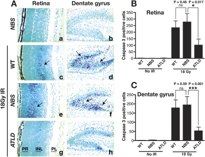
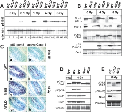
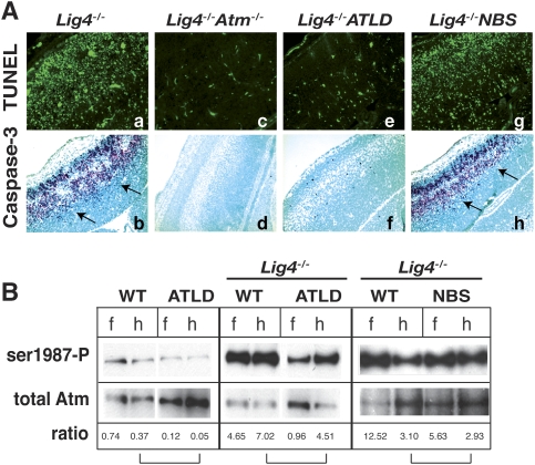
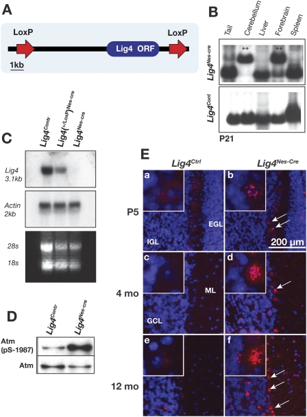
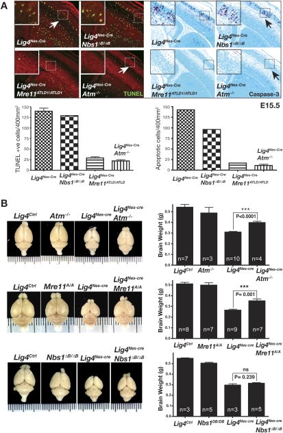
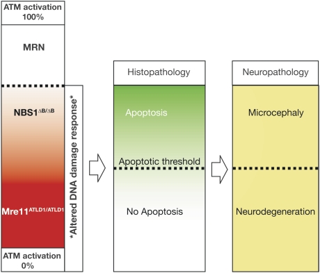
The MRN complex (Mre11/RAD50/NBS1) and ATM (ataxia telangiectasia, mutated) are critical for the cellular response to DNA damage. ATM disruption causes ataxia telangiectasia (A-T), while MRN dysfunction can lead to A-T-like disease (ATLD) or Nijmegen breakage syndrome (NBS). Neuropathology is a hallmark of these diseases, whereby neurodegeneration occurs in A-T and ATLD while microcephaly characterizes NBS. To understand the contrasting neuropathology resulting from Mre11 or Nbs1 hypomorphic mutations, we analyzed neural tissue from Mre11(ATLD1/ATLD1) and Nbs1(DeltaB/DeltaB) mice after genotoxic stress. We found a pronounced resistance to DNA damage-induced apoptosis after ionizing radiation or DNA ligase IV (Lig4) loss in the Mre11(ATLD1/ATLD1) nervous system that was associated with defective Atm activation and phosphorylation of its substrates Chk2 and p53. Conversely, DNA damage-induced Atm phosphorylation was defective in Nbs1(DeltaB/DeltaB) neural tissue, although apoptosis occurred normally. We also conditionally disrupted Lig4 throughout the nervous system using Nestin-cre (Lig4(Nes-Cre)), and while viable, these mice showed pronounced microcephaly and a prominent age-related accumulation of DNA damage throughout the brain. Either Atm-/- or Mre11(ATLD1/ATLD1) genetic backgrounds, but not Nbs1(DeltaB/DeltaB), rescued Lig4(Nes-Cre) microcephaly. Thus, DNA damage signaling in the nervous system is different between ATLD and NBS and likely explains their respective neuropathology.


Figures

Figure 1.
Figure 1.
Radiation-induced apoptosis in Mre11ATLD1/ATLD1 and Nbs1ΔB/ΔB CNS. (A) Wild-type (WT), Mre11ATLD1/ATLD1, and Nbs1ΔB/ΔB mice at P5 were treated with 8Gy of γ-irradiation (panels c–h) and collected 6 h post-IR. Nonirradiated sections are shown in panels a and b. Immunostaining was performed against active-caspase 3. (Panels a,c,e,g) Images of the retina are at 400× magnification. (Panels b,d,f,h) Images of the dentate gyrus are at 200× magnification. Retinal photoreceptor layer (PR), inner nuclear layer (INL), and plexiform layer (PL) are indicated. Arrows point to immunopositive cells. The number of activated caspase-3-positive cells in the retina (B) or dentate gyrus (C) were quantified; error bars represent standard deviation.
Figure 2.
Figure 2.
DNA damage responses in the Mre11ATLD1/ATLD1 and Nbs1ΔB/ΔB nervous system at postnatal day 5. (A) Wild-type (WT), Mre11ATLD1/ATLD1, and Nbs1ΔB/ΔB mice were treated with indicated doses (Gy) of IR, and cerebella were harvested 30 min post-IR. Total Atm was immunoprecipitated from cerebellar lysates followed by immunoblotting for Atm pSer1987. Total Atm served as the loading control. Ratios represent the relative increase of phosphorylated Atm compared with total Atm protein. (B) Wild-type (WT), Mre11ATLD1/ATLD1, and Nbs1ΔB/ΔB mice were treated with IR and cerebella were harvested 3 h post-IR. Cerebellar lysates were probed using antibodies recognizing Nbs1, Mre11, and Chk2. Phosphorylated Chk2 (pChk2) was detected as an upward mobility shift. β-actin was used as a loading control. (C) Immunostaining 1 h post-IR (18 Gy) of wild-type, Mre11ATLD1/ATLD1, and Nbs1ΔB/ΔB mice to detect pSer18 of p53 (p53-ser18) and active-caspase-3 (200× magnification). (D) Wild type (WT), Atm−/−, and Mre11ATLD1/ATLD1 were treated with 4 Gy of IR to determine comparative Chk2 and p53 phosphorylation after 1 h in Atm−/− cerebellum. (E) Wild-type (WT), Mre11ATLD1/ATLD1, and Nbs1ΔB/ΔB mice were treated with 18 Gy of IR and collected 3 h post-IR. Cerebellar lysates in D and E were probed for Chk2, p53 (pSer18), and Nbs1 or Mre11. Ponceau staining is shown to indicate relative protein loading.
Figure 3.
Figure 3.
DNA damage signaling in ATLD and NBS after Lig4 loss. (A) TUNEL analysis and immunostaining against active caspase-3 was performed on E15.5 cryosections of Lig4−/−, Lig4−/−;Atm−/−, Lig4−/−;Mre11ATLD1/ATLD1, and Lig4−/−;Nbs1ΔB/ΔB. Arrows indicate immunopositive signal. Images of the neopallial cortex were captured at a 200× magnification. (B) Tissue lysates were prepared from E13.5 forebrain (f) and hindbrain (h) collected from the indicated genotypes. Total Atm was immunoprecipitated from lysates followed by immunoblotting for phosphorylated Atm (pser1987). Quantitative analysis depicted below the blot indicates the relative ratio between phospho ser1987 and total Atm; brackets indicate matched brain regions.
Figure 4.
Figure 4.
Generation and analysis of conditional Lig4-null mice. (A) The conditional Lig4 allele was generated by flanking the Lig4 ORF by LoxP sites. (B) Deletion of the Lig4 allele throughout the brain was done using Nestin-cre. Southern blot analysis of DNA from 1-mo-old Lig4Nes-Cre or Lig4Cont mice shows the deleted Lig4 allele (1.6 kb) in cerebellum and forebrain (**) but not in liver or spleen. (C) RNA was extracted from adult cerebella for Northern analysis. A full-length Lig4 cDNA probe was used to detect expression of Lig4 mRNA. (D) Cerebella were collected from P5 Lig4Nes-Cre and Lig4Ctrl animals. Atm was immunoprecipitated from tissue lysates, then immunoblotted for Atm pSer1987 and total Atm. (E) Lig4Nes-Cre and Lig4Ctrl tissues were collected at the indicated ages and immunostained for γH2AX. Representative images of cerebella from each age and genotype are shown (200× magnification). Arrows indicate that after Lig4 deletion Purkinje cells contain γH2AX; inset panels show Purkinje cells with γH2AX foci at 800× magnification.
Figure 5.
Figure 5.
Impact of ATLD and NBS mutations in Lig4Nes-Cre. (A) TUNEL analysis and immunostaining for active caspase-3 were performed on E15.5 embryos from indicated genotypes. The total number of positive cells per 400 μm2 was quantified; error bars represent standard deviation. Micrographs encompass ganglionic eminence and neopallial cortex (200× magnification). (B) The effect of A-T, ATLD, and NBS mutations on Lig4Nes-Cre brain size. Brain weights were collected from sex-matched cohorts at 4 mo of age. No significant difference was observed between Lig4Ctrl groups and Atm−/− or Mre11ATLD1/ATLD1 controls; however a statistically significant decrease in Nbs1ΔB/ΔB brain weight was observed compared with controls (P = 0.013). A statistically significant recovery in Lig4Nes-Cre brain weight was observed in Lig4Nes-Cre;Atm−/− and Lig4Nes-Cre;Mre11ATLD1/ATLD1 but not in Lig4Nes-Cre;Nbs1ΔB/ΔB mice. Error bars represent standard deviation.
Figure 6.
Figure 6.
Neuropathology in ATLD and NBS. Full ATM activation and activity require the MRN complex. Both Mre11ATLD1/ATLD1 and Nbs1ΔB/ΔB mice show defective Atm phosphorylation. However, Atm activity is substantially higher in the Nbs1ΔB/ΔB nervous system compared with the Mre11ATLD1/ATLD1 nervous system and is sufficient to activate apoptosis. The different apoptotic response observed between the Mre11ATLD1/ATLD1 and Nbs1ΔB/ΔB mice suggests a threshold level of Atm activity is required to induce apoptosis during neurogenesis. In the human brain, NBS mutations elevate DNA damage, leading to increased apoptosis and microcephaly. In contrast, ATLD mutations cannot sufficiently activate ATM, thereby failing to engage apoptosis and eliminate DNA damaged cells. Similar to A-T, damaged cells may become incorporated into the nervous system and at later times will malfunction and die, resulting in progressive neurodegeneration.

References

    1. Ahn J.Y., Schwarz J.K., Piwnica-Worms H., Canman C.E. Threonine 68 phosphorylation by ataxia telangiectasia mutated is required for efficient activation of Chk2 in response to ionizing radiation. Cancer Res. 2000;60:5934–5936. - PubMed
    1. Barnes D.E., Stamp G., Rosewell I., Denzel A., Lindahl T. Targeted disruption of the gene encoding DNA ligase IV leads to lethality in embryonic mice. Curr. Biol. 1998;8:1395–1398. - PubMed
    1. Bredemeyer A.L., Sharma G.G., Huang C.Y., Helmink B.A., Walker L.M., Khor K.C., Nuskey B., Sullivan K.E., Pandita T.K., Bassing C.H., et al. ATM stabilizes DNA double-strand-break complexes during V(D)J recombination. Nature. 2006;442:466–470. - PubMed
    1. Buis J., Wu Y., Deng Y., Leddon J., Westfield G., Eckersdorff M., Sekiguchi J.M., Chang S., Ferguson D.O. Mre11 nuclease activity has essential roles in DNA repair and genomic stability distinct from ATM activation. Cell. 2008;135:85–96. - PMC - PubMed
    1. Callen E., Jankovic M., Difilippantonio S., Daniel J.A., Chen H.T., Celeste A., Pellegrini M., McBride K., Wangsa D., Bredemeyer A.L., et al. ATM prevents the persistence and propagation of chromosome breaks in lymphocytes. Cell. 2007;130:63–75. - PubMed

Publication types

MeSH terms