. 2009 Apr 30;113(18):4240-9.
doi: 10.1182/blood-2008-10-183251. Epub 2009 Jan 26.

IL-17-producing human peripheral regulatory T cells retain suppressive function

Affiliations

IL-17-producing human peripheral regulatory T cells retain suppressive function

Gaëlle Beriou et al. Blood. .

Abstract

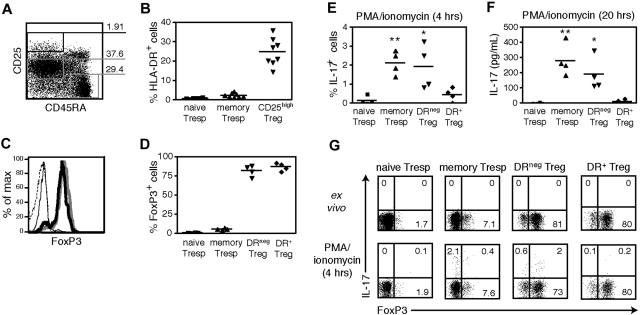
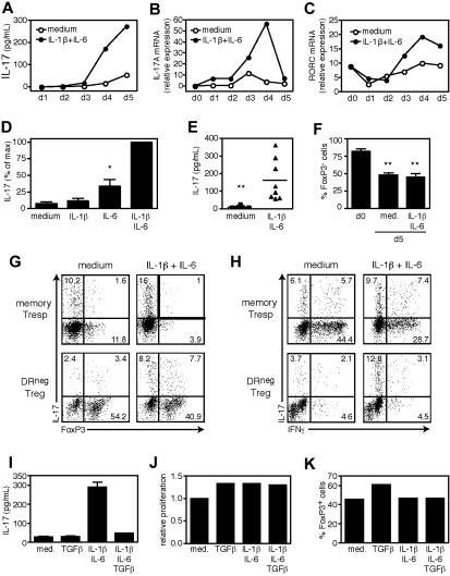
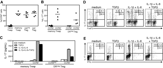
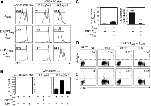
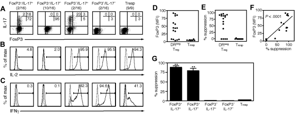
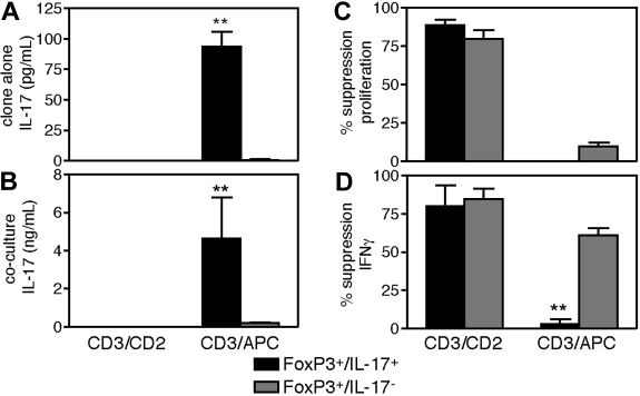
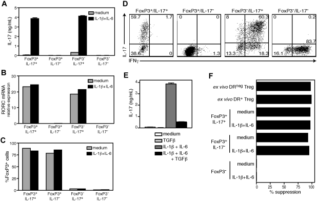
Although implicated in antagonistic functions, both regulatory T cells (Tregs) and Th17 effector cells play an important role in controlling autoimmune pathogenesis. Paradoxically, recent studies indicate that Tregs have the capacity to produce interleukin-17 (IL-17), although the ability of these cells to retain their suppressive function remains unknown. Here we report that human Tregs within the CD4(+)CD45RA(-)CD25(high)CCR6(+)HLA-DR(-)FoxP3(+) population produce IL-17 when activated in the presence of the proinflammatory cytokines IL-1beta and IL-6, whereas IL-17 secretion was inhibited by TGFbeta. To assess the ability of a single Treg to secrete IL-17 and to suppress in vitro immune function, we isolated clones from this population. We found that IL-17(+)/FoxP3(+) Treg clones retain suppressive function and exhibit the plasticity to secrete IL-17 or suppress depending on the nature of the stimulus provided. IL-17 production by these Treg clones was accompanied by sustained FoxP3 expression and concomitant, but reversible, loss of suppressive activity. Our data demonstrate that at the single cell level a subset of in vitro suppressive FoxP3(+) cells can be driven to secrete IL-17 under inflammatory conditions. These findings suggest a new mechanism by which inflammation can drive Tregs to secrete IL-17, thereby dampening suppression and promoting an inflammatory milieu.


Figures

Figure 1
Figure 1
Sorted DR Tregs, but not DR+ Tregs, produce IL-17 in response to short-term mitogenic activation. (A) Gating strategy for FACS sorting of CD4+ peripheral blood T cells into populations of Tregs (CD45RACD25high), memory Tresps (CD45RACD25med), and naive Tresps (CD45RA+CD25). (B) Percentages of HLA-DR+ cells within the 3 different gates (n = 9). (C) FoxP3 intracellular staining of FACS-sorted naive Tresps (dashed line), memory Tresps (thin solid line), DR Tregs (bold line), and DR+ Tregs (gray line). (D) The percentages of FoxP3+ cells is shown (n = 4). (E) FACS-sorted naive Tresps, memory Tresps, DR Tregs, and DR+ Tregs (104 cells/well) were stimulated in serum-free X-Vivo medium for 4 hours with PMA/ionomycin and GolgiStop, and stained for intracellular IL-17 (n = 4). (F) IL-17 levels measured by ELISA in the same samples after 20 hours of stimulation with PMA/ionomycin. (G) One representative staining of intracellular FoxP3 and IL-17 in the 4 populations ex vivo or after 4 hours of stimulation with PMA/ionomycin and GolgiStop. **P < .01; *P < .05.
Figure 2
Figure 2
IL-17 secretion by DR Tregs is induced by IL-1β and IL-6 and inhibited by TGFβ. FACS-sorted DR Tregs (104 cells/well) were stimulated in serum-free X-Vivo medium for 5 days with plate-bound αCD3, soluble αCD28, and IL-2 in the presence of exogenous IL-1β and IL-6. Supernatants and cells were harvested at 24-hour intervals and analyzed for IL-17 content by ELISA (A) or RNA expression of IL-17A (B) and RORC (C) by real-time polymerase chain reaction (PCR). Data are representative of 2 independent experiments. (D) IL-17 secretion measured by ELISA in day 5 supernatants and represented as percentage of max (n = 5). (E) The IL-17 levels in day 5 supernatants are shown (n = 8). (F) Percentages of FoxP3+ cells ex vivo (n = 4) or on day 5 of stimulation (n = 8). At the end of the culture, cells were pulsed for 4 hours with PMA/ionomycin and GolgiStop, and stained for intracellular IL-17 versus FoxP3 (G) or IFNγ (H). Data are representative of 4 independent experiments. (I) DR Tregs were stimulated for 5 days in the presence of exogenous IL-1β/IL-6 or TGFβ and tested for IL-17 secretion by ELISA. (J) Although the cultures were established at identical cell numbers, the relative cell numbers at the end of culture were determined for each condition by flow cytometry and represented as relative proliferation versus untreated cells. (K) Percentages of FoxP3+ cells on day 5 of stimulation. Data are representative of 5 independent experiments. **P < .01; *P < .05.
Figure 3
Figure 3
The DR Tregs that produce IL-17 express CCR6 ex vivo. (A) Percentages of CCR6+ cells in gated ex vivo naive Tresps (n = 4), memory Tresps (n = 4), DR Tregs (n = 6) and DR+ Tregs (n = 6). (B) CCR6 and CCR6+ fractions of DR Tregs or memory Tresps were FACS-sorted and stimulated for 5 days with αCD3/αCD28 and IL-2 in the presence of IL-1β/IL-6. The percentages of IL-17+ cells at the end of the culture is shown (n = 4). (C) Cells were stimulated in the presence of IL-1β/IL-6 or TGFβ, and analyzed for IL-17 secretion by ELISA. At the end of the culture, CCR6+DR Tregs were stained for intracellular IL-17 versus FoxP3 (D) or IFNγ (E). Data are representative of 4 independent experiments. **P < .01; *P < .05.
Figure 4
Figure 4
IL-17 secretion upon coculture of DR Tregs with Tresp cells inversely correlates with suppression. (A) CFSE-labeled CD25 Tresp cells were cultured in RPMI medium containing 5% human AB serum, alone or with DR Treg or DR+ Treg (1:1 ratio) under 3 different stimulatory conditions: αCD3/αCD2 beads (left), or irradiated T-cell depleted APCs and plate-bound αCD3 at 0.1 μg/mL (middle) or 0.5 μg/mL (right). On day 4 of stimulation, proliferation of Tresp cells was analyzed by assessing CFSE dilution by FACS analysis. The numbers represent the percentage of Tresp cells that divided (CFSE dilution). (B) IL-17 production in these differentially stimulated Tresp cultures or DR or DR+ Treg cocultures was analyzed by ELISA from day 4 supernatants. Data shown are representative of 4 independent experiments. (C) The results of the same assay performed on 4 independent donors and represented as percentage suppression of proliferation and fold induction of IL-17 in DR and DR+ Treg cocultures stimulated with αCD3 (0.5 μg/mL) and APCs (mean + SEM). (D) Cultures containing CFSE-labeled Tresp cells, DR Tregs, or both cell types (1:1 ratio) were stimulated with high dose αCD3 and APCs. On day 4, PMA/ionomycin and GolgiStop were added to the cultures for a 4-hour pulse before the cells were harvested, surface stained with αCD2 to distinguish the CD4 T cell populations from the irradiated, T-depleted APCs, and then permeabilized and stained for IL-17. Data shown are representative of 3 independent experiments. **P < .01.
Figure 5
Figure 5
One subset of DR Treg clones can suppress or secrete IL-17 in response to different stimuli. The clones that grew from wells seeded at 1 DR Treg per well were analyzed for several features and compared with clones derived from Tresp cells. After 5 weeks of expansion, a portion of each clone was stained for FoxP3 and IL-17 expression and tested for ability to suppress the proliferation of freshly isolated Tresp cells in cocultures stimulated with αCD3/αCD2 beads. On day 4, half the media in each well replaced with 3H thymidine to monitor proliferation. Data are representative of 2 independent cloning experiments. Intracellular expression of FoxP3/IL-17 (A), IL-2 (B), and IFNγ (C) of DR Treg clones after 4 hours of stimulation with PMA/ionomyicin and GolgiStop. One representative clone from each pattern is shown. (D) Mean FoxP3 expression by each clone. (E) The suppressive capacity of each clone represented as percentage suppression. (F) Mean FoxP3 expression by each clone is shown relative to its suppressive ability. (G) Suppression by each pattern of clone.
Figure 6
Figure 6
IL-17 production by FoxP3+/IL-17+ clones results in concomitant loss of suppressive function. The suppressive and effector functions of the clones were assessed in cocultures stimulated with αCD3/αCD2 beads or with 0.5 μg/mL αCD3 and APCs. On day 4, half the media in each well was removed, interrogated for content of IL-17 and IFNγ by ELISA, and replaced with 3H thymidine to monitor proliferation. Two representative clones for each pattern are shown. Data are representative of 2 independent cloning experiments. Panels show the ability of the clones to secrete IL-17 in response to the different stimuli (A), the levels of IL-17 induced in the respective clone cocultures (B), the ability of the clones to suppress Tresp proliferation (C), and the ability of the clones to suppress IFNγ production (D). **P < .01.
Figure 7
Figure 7
FoxP3+/IL-17+ clones remain suppressive after they have been induced to secrete IL-17. The clones derived from the DR Treg population were stimulated for 5 days with αCD3/αCD28 and IL-2 in the presence of IL-1β/IL-6, and analyzed for IL-17 secretion by ELISA (A), RORC mRNA expression by real-time PCR (B), and FoxP3 expression by intracellular staining (C). (D) Intracellular IL-17/IFNγ staining of IL-1β/IL-6 treated clones. (E) FoxP3+/IL-17+ clones were stimulated in the presence of IL-1β/IL-6 or TGFβ, and analyzed for IL-17 secretion by ELISA. (F) IL-1β/IL-6 treated clones were expanded for 2 weeks and tested for suppressive function in cocultures stimulated with αCD3/αCD2 beads, as compared with freshly isolated DR and DR+ Tregs. One representative clone from each pattern is shown.

References

    1. Thornton AM, Shevach EM. CD4+CD25+ immunoregulatory T cells suppress polyclonal T-cell activation in vitro by inhibiting interleukin 2 production. J Exp Med. 1998;188:287–296. - PMC - PubMed
    1. Baecher-Allan C, Brown JA, Freeman GJ, Hafler DA. CD4+CD25high regulatory cells in human peripheral blood. J Immunol. 2001;167:1245–1253. - PubMed
    1. Levings MK, Sangregorio R, Roncarolo MG. Human cd25(+)cd4(+) t regulatory cells suppress naive and memory T-cell proliferation and can be expanded in vitro without loss of function. J Exp Med. 2001;193:1295–1302. - PMC - PubMed
    1. Jonuleit H, Schmitt E, Stassen M, Tuettenberg A, Knop J, Enk AH. Identification and functional characterization of human CD4(+)CD25(+) T cells with regulatory properties isolated from peripheral blood. J Exp Med. 2001;193:1285–1294. - PMC - PubMed
    1. Dieckmann D, Plottner H, Berchtold S, Berger T, Schuler G. Ex vivo isolation and characterization of CD4(+)CD25(+) T cells with regulatory properties from human blood. J Exp Med. 2001;193:1303–1310. - PMC - PubMed

Publication types

MeSH terms