. 2009 Feb 3;106(5):1462-7.
doi: 10.1073/pnas.0812017106. Epub 2009 Jan 26.

Expression profiling during ocular development identifies 2 Nlz genes with a critical role in optic fissure closure

Affiliations

Expression profiling during ocular development identifies 2 Nlz genes with a critical role in optic fissure closure

Jacob D Brown et al. Proc Natl Acad Sci U S A. .

Abstract

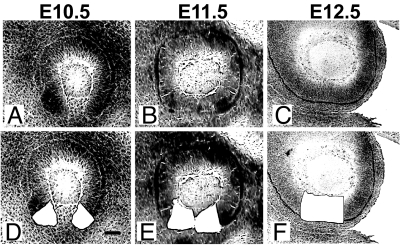
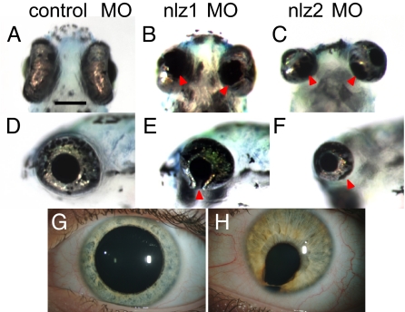
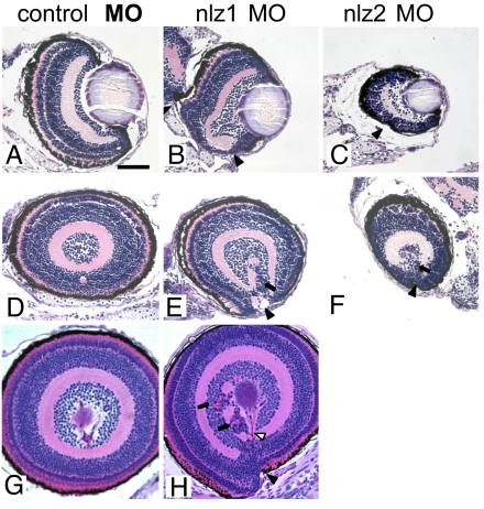
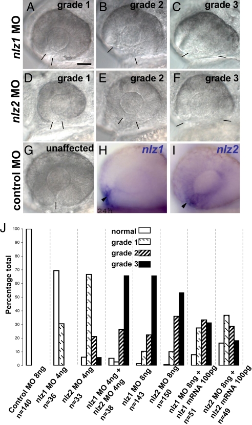
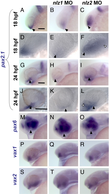
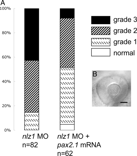
The gene networks underlying closure of the optic fissure during vertebrate eye development are poorly understood. Here, we profile global gene expression during optic fissure closure using laser capture microdissected (LCM) tissue from the margins of the fissure. From these data, we identify a unique role for the C(2)H(2) zinc finger proteins Nlz1 and Nlz2 in normal fissure closure. Gene knockdown of nlz1 and/or nlz2 in zebrafish leads to a failure of the optic fissure to close, a phenotype which closely resembles that seen in human uveal coloboma. We also identify misregulation of pax2 in the developing eye of morphant fish, suggesting that Nlz1 and Nlz2 act upstream of the Pax2 pathway in directing proper closure of the optic fissure.


Conflict of interest statement

The authors declare no conflict of interest.

Figures

Fig. 1.
Fig. 1.
Isolation of optic fissure tissue and experimental design. Representative sagittal sections through the eye of wild-type C57BL6/J mice at E10.5 (A and D), E11.5 (B and E), and E12.5 (C and F). Hematoxylin stained sections are seen before (A–C) and after (D–F) laser capture microdissection. Isolated tissue was used in expression profiling experiment. (Scale bar, 50 μm.)
Fig. 2.
Fig. 2.
Eye phenotype in nlz1 and nlz2 morphant zebrafish. At 5 dpf, fish injected with control MO show a completely fused optic fissure in the ventral eye (A, ventral; D, lateral) whereas there is an obvious coloboma in the ventral eye of the nlz1 (B, ventral; E, lateral) and nlz2 (C, ventral; F, lateral) morphant fish indicated at arrowheads. This phenotype recapitulates human uveal coloboma (H). Unaffected human eye (G). (Scale bar, 200 μm.)
Fig. 3.
Fig. 3.
Histopathology of nlz morphant fish. Coronal (A–C) and sagittal (D–H) planes through the zebrafish eye. Normal retinal lamination and a fused ventral fissure can be seen in control MO injected fish at 5 dpf (A and D) and at 6 dpf (G) (Top, dorsal; Bottom, ventral). Fissure closure defects can be seen in nlz1 morphant fish at 5 dpf (B and E) and 6 dpf (H) and nlz2 morphant fish at 5 dpf (C and F). The coloboma in morphant fish is accompanied by discontinuous RPE (black arrowhead), retinal dysplasia/rosettes (arrows), and abnormal vasculature (open arrowhead). H&E staining.
Fig. 4.
Fig. 4.
Phenotypic grades of nlz morphant fish 24 hpf. nlz1 MO (A–C) or nlz2 MO (D–F) splice site MO injected fish were scored as having a grade 1 (A and D), grade 2 (B and E), or grade 3 (C and F) optic fissure defect. At 24 hpf, the margins of the optic fissure are apposed in normal eyes (G) and closure is just beginning. Bars delineate edges of optic fissure. (Scale bar, 50 μm.) The results of the phenotype profiling are summarized in the bar graph (J) where lower grade closure defects are observed at low doses of either nlz1 or nlz2 MO, yet an effect which is at least additive is seen when these doses are combined. A higher dose of either morpholino produces strong optic fissure defects in morphant fish, which is partially rescued by coinjection of respective wild-type mRNA. Expression of nlz1 (H) and nlz2 (I) in wild-type zebrafish embryos at 24 hpf shows expression at the optic fissure (arrowheads).
Fig. 5.
Fig. 5.
Misregulation of pax2.1 and pax6 in nlz morphant fish in the setting of normal vax1 and vax2 expression at 24 hpf. In situ hybridization at 18 hpf (A–F) and 24 hpf (G–U). At 18 hpf, pax2.1 is normally expressed in the optic stalk and ventral optic primordium (A and D, arrowheads) and at 24 hpf is expressed distinctly in the optic stalk and fissure (G and J, arrowheads), midbrain/hindbrain boundary, and otic vesicle. In nlz1 morphant fish, pax2.1 expression is drastically reduced in the optic stalk and fissure (B, E, H, and K). In nlz2 morphant fish, the expression domain is expanded (C, F, I, L, open arrowheads). A higher magnification view shows the expression of pax2.1 in normal and nlz morphant fish at 18 hpf (D–F) and 24 hpf (J–L). (M–O) pax6 expression shows a subtle increase in the ventral eye in nlz1 morphant fish (N) and slight decrease in nlz2 fish (O, arrowheads). vax1 (P–R) and vax2 (S–U) are both expressed at normal levels and in a normal distribution in the optic stalk and ventral retina. (Scale bar, 100 μm.)
Fig. 6.
Fig. 6.
pax2.1 partially rescues nlz1 morphant phenotype. (A) Coinjection of pax2.1 mRNA shifts the coloboma phenotype profile toward normal, decreasing the percentage of embryos in cohort with severe defects (grade 3) and increasing the percentage of mildly affected (grade 1) and normal eyes. (B) Representative picture of nlz1 morphant eye at 24 hpf rescued by pax2.1 injection.
Fig. 7.
Fig. 7.
NLZ1 and NLZ2 bind the PAX2 promoter. Chromatin immunoprecipitation with anti-FLAG Ab shows FLAG-tagged NLZ1 and NLZ2 bind to a conserved 123-bp region of the PAX2 promoter containing 1 putative zinc finger binding site (see also Fig. S5).

References

    1. O'Rahilly The early development of the eye in staged human embryos. Contrib Embryol. 1966;XXXVIII(25–263):1–45.
    1. Bermejo E, Martinez-Frias ML. Congenital eye malformations: clinical-epidemiological analysis of 1,124,654 consecutive births in Spain. Am J Med Genet. 1998;75(5):497–504. - PubMed
    1. Porges Y, et al. Hereditary microphthalmia with colobomatous cyst. Am J Ophthalmol. 1992;114(1):30–34. - PubMed
    1. Maumenee IH, Mitchell TN. Colobomatous malformations of the eye. Trans Am Ophthalmol Soc. 1990;88:123–132. discussion 133–125. - PMC - PubMed
    1. Traboulsi E. Colobomatous micropthalmia, anophthalmia, and associated malformation syndromes. In: Traboulsi E, editor. Genetic Diseases of the Eye. New York: Oxford Univ Press; 1999. pp. 51–80.

Publication types

Associated data